So what’s the story with antioxidants? Do they make a difference or not? If you listen to the buzz, you end up more confused than ever.
- According to Julian Whitaker, M.D., “The jury has reached a verdict and I urge my medical colleagues to listen carefully. It’s no longer a question of should our patients be taking antioxidant supplements, but rather which ones and why. It’s time that physicians recognize the value of the growing body of research showing the many health benefits of natural antioxidants.”
- But if you listen to Stephen Barrett, M.D. at Quackwatch (for a contrary perspective), “There is widespread scientific agreement that eating adequate amounts of fruits and vegetables can help lower the incidence of cardiovascular disease and certain cancers. With respect to antioxidants and other phytochemicals, the key question is whether supplementation has been proven to do more good than harm. So far, the answer is no, which is why the FDA will not permit any of these substances to be labeled or marketed with claims that they can prevent disease.”1 Stephen Barrett, M.D. “Antioxidants and Related Phytochemicals: Current Scientific Perspective.” Quackwatch November 14, 2012. (Accessed 28 Jan 2015.) http://www.quackwatch.org/03HealthPromotion/antioxidants.html
- Or this from the Professor Tony Segal of the University College of London’s Centre for Molecular Medicine, “Many patients might be using expensive antioxidant drugs based upon completely invalid theories as to their therapeutic potential.”2 “UCL study questions basis for treatment of diseases including cancer and arthritis” UCL News 26 February 2004. (Accessed 29 Jan 2015.) http://www.ucl.ac.uk/news/news-articles/news-releases-archive/oxygen
- Confused, yet? Well then, how about this from The Rotterdam Study of Dietary antioxidants and Parkinson disease. “CONCLUSION: Our data suggest that a high intake of dietary vitamin E may protect against the occurrence of Parkinson’s disease.”3 de Rijk MC, Breteler MM, et al. “Dietary antioxidants and Parkinson disease. The Rotterdam Study.” Arch Neurol. 1997 Jun;54(6):762-5. http://www.ncbi.nlm.nih.gov/pubmed/9193212
Get the idea? There is a great deal of confusion and contradictory information out there on antioxidants. So let’s try and clear some of it up.
What Exactly Is a Free Radical?
A free radical is an especially reactive atom or group of atoms that has one or more unpaired electrons. If you remember your high school chemistry, you will remember that electrons come in pairs. When one electron is lost from that pair, it makes the atom “highly reactive” as it looks to replace that lost electron anywhere it can. In your body, those replacement electrons come from cells in your body–destroying those cells in the process. Free radicals put your body in a state of oxidative stress in which your body is no longer able to maintain a balance between the appearance of reactive oxygen species and its ability to detoxify those free radicals or to repair the resulting damage. That’s why free radicals function as cellular killers that wreak havoc by damaging DNA, altering biochemical compounds, corroding cell membranes, and destroying cells outright.
Scientists now know that free radicals play a major role in the aging process as well as in the onset of cancer, heart disease, stroke, arthritis, and possibly allergies and a host of other ailments. The link between free radicals and the “aging diseases” is the most important discovery since doctors learned that some illnesses are caused by germs.
In a very real sense, the free radical process in our bodies is much the same as the process that causes fuel to burn and oil to go rancid or an apple to turn brown if you slice it open and expose it to air. It is as though our bodies rust from the inside out. When the process gets really out of control, it can cause tumors, hardening of the arteries, and macular degeneration — not to mention wrinkled skin — to name just a few of the negative effects.
Another way to think of free radicals is as ravenous molecular sharks–sharks so hungry that in little more than a millionth of a second, they can be making a frenzied attack on a healthy neighboring cellular molecule. A single free radical can destroy an enzyme, a protein molecule, a strand of DNA, or an entire cell. Even worse, that one free radical can unleash, in a fraction of a second, a torrential chain reaction that produces a million or more additional killer free radicals–each out hunting for living cells to destroy like a herd of sharks in a mindless feeding frenzy.
Free Radicals and Cancer
The connection between free radicals and cancer has been recognized for decades, but the underlying mechanisms have steadily become more apparent. For example, recently, it is becoming increasingly clear that metabolic oxidative free radical reactions are altered in cancer cells vs. normal cells. The current consensus is that cancer cells may exist in a chronic state of metabolic oxidative stress which may represent a significant underlying mechanism contributing to that cell’s malignancy. A unifying goal of the investigators in the Free Radical Cancer Biology Program, out of the University of Iowa Hospitals & Clinics, is to utilize a comprehensive understanding of redox biology to develop new biochemical techniques for improving cancer therapy by taking advantage of fundamental differences in the oxidative metabolism between cancerous cells and normal cells.4 “Free Radical Cancer Biology Program.” University of Iowa Hospitals & Clinics. (Accessed 29 Jan 2015.) http://www.uihealthcare.org/otherservices.aspx?id=21027
 There are four primary sources of free radicals:
- The Environment. Air pollution, cigarette smoke, smog, soot, automobile exhaust, toxic waste, pesticides, herbicides, ultraviolet light, background radiation, drugs, and even certain foods can all generate free radicals in the body.
- Internal Production. Our bodies constantly produce free radicals as a byproduct of normal metabolism. And yes, since exercise increases metabolic rates, it increases the production of free radicals.
- Stress Factors. Aging, trauma, medications, disease, infection, and “stress” itself all accelerate the body’s production of free radicals — by a factor of eight, or more.
- Chain Reactions. When a free radical steals an electron to balance itself, it creates a new free radical in the molecule from which it stole the electron. In many cases, the new free radical will seek to balance itself by stealing its own electron — and on and on. And remember, even one free radical is capable of destroying an entire cell, or a strand of DNA. Imagine what a chain reaction of millions of free radicals can do!
Types of Free Radicals
There are different types of free radicals in the body. They each work in different areas of the body and on different cells or even different parts of cells. Four particularly destructive ones are:
- Superoxide Radical. This radical tries to steal its much-needed electron from the mitochondria of the cell. When mitochondria are destroyed, the cell loses its ability to convert food to energy. It dies.
- Hydroxyl radical. This free radical attacks enzymes, proteins, and the unsaturated fats in cell membranes.
- Lipid Peroxyl Radical. This radical unleashes a chain reaction of chemical events that can so totally compromise the cellular membrane that the cell bursts open, spews its contents, and dies.
- Singlet Oxygen. Not technically a free radical, this metabolite can nevertheless wreak havoc on the body. It’s listed here because it functions like a free radical and because it is controlled by antioxidants.
Antioxidants to the Rescue
Your body is constantly replacing and repairing free-radical damaged cells; but with the way we live and abuse ourselves, our bodies are bombarded with more free radicals than they can handle. By supplementing with antioxidants, we help our bodies keep up with the carnage. It is even possible to get ahead of the game and reverse some of that damage. So what are antioxidants?
Antioxidants, quite simply, are compounds that render free radicals harmless and stop the chain reaction formation of new free radicals. There are three sources of antioxidants. First, several metabolic enzymes produced by the body–glutathione peroxidase, superoxide dismutase, and catalase–are extremely effective antioxidant scavengers.5 Etienne Pigeolet, Philippe Corbisier, et al. “Glutathione peroxidase, superoxide dismutase, and catalase inactivation by peroxides and oxygen derived free radicals.” Mech Ageing Dev. 1990 Feb 15;51(3):283-97. http://www.ncbi.nlm.nih.gov/pubmed/2308398 Unfortunately, the body’s ability to produce these enzymes fades dramatically in our late twenties. Second, many foods and plants provide powerful antioxidants. Among these are vitamins E and C and beta carotene (but only in their natural full-complex forms) and the proanthocyanidins (including grape seed and green tea extract). And finally, cutting edge research is continually uncovering new antioxidants in foods and plants from around the world–as well as synthesizing them in the laboratory.
In summary, many scientists now believe that free radicals are the major villain in both aging and the diseases associated with aging. The amount of cells destroyed over the years by free radicals is enormous. Free radicals literally “eat away” the major organs of the body. The use of antioxidant supplements at a maintenance level may provide the ultimate defense against premature aging and a compromised immune system. At therapeutic levels, antioxidants may actually play a significant role in reversing many of the effects of aging and disease.
But It’s More Complicated than that
When it comes to free radicals and antioxidants, it’s not a simple case of black and white. The shades of gray in this discussion are very important. For example, although free radicals likely play a major role in the onset of many diseases including cancer and antioxidants offer protection, things change somewhat if your immune system is under attack or if you actually get cancer. Then, in some cases, the roles reverse. Take the superoxide radical we mentioned earlier. Superoxide is biologically quite toxic and extremely harmful to healthy cells. However, it is also deployed by the immune system to kill invading microorganisms. In phagocytes, superoxide is produced in large quantities by the enzyme NADPH oxidase for use in oxygen-dependent killing mechanisms of invading pathogens. If that system is not working properly, you have a problem. For example, mutations in the gene coding for the NADPH oxidase enzyme complex means your immune system can’t produce the superoxide radical. This results in an immunodeficiency syndrome called chronic granulomatous disease, which is characterized by extreme susceptibility to infection, especially catalase positive organisms.6 B H Segal, P Veys, H Malech, M J Cowan. “Chronic Granulomatous Disease: Lessons from a Rare Disorder.” Biol Blood Marrow Transplant. Jan 2011; 17(1 Suppl): S123–S131. http://www.ncbi.nlm.nih.gov/pmc/articles/PMC3052948/ To put this in simpler terms: white blood cells produce oxygen free radicals, and the process by which they do so is essential for the efficient killing of microbes. In this particular instance, free radicals are not bad; they’re an essential part of your immune system and good health.
When it comes to cancer, we see the same dichotomy. In fact, it has been recognized that the primary medical therapies used to treat cancer–radiation and chemotherapy–work by causing oxidative stress that ultimately kills cancer cells. The idea is that by their aggressive nature, cancer cells suck up more of the free radicals than healthy cells, thus, they die first–hopefully–allowing the healthy cells to survive and recover. This is the reason many oncologists insist that you stop taking any antioxidants while on a chemotherapy regimen so as not to undermine the treatment. In fact, there are indications that your immune system uses the same strategy in certain case, just more targeted, when fighting cancer. In fact, some researchers are going so far as to say that genetic or pharmacologic inhibition of antioxidant proteins might be a useful therapeutic approach for dealing with cancer in humans.7 Elizabeth G. Phimister, Navdeep S. Chandel, David A. Tuveson. “The Promise and Perils of Antioxidants for Cancer Patients.” NEJM, 2014; 371 (2): 177. http://www.nejm.org/doi/full/10.1056/NEJMcibr1405701
However, there is no absolute rule here as some antioxidants aggressively attack cancer cells themselves. So, what do you do? Well, to paraphrase Kenny Rogers, “When it comes to free radicals and antioxidants, you have to know when to hold them and know when to fold them. You have to know when to take your supplements and know when to run.”
So Why the Inconsistent Test Results?
Now that we have a little understanding as to what antioxidants are and how they work, let’s go back to where we started and see if we can understand why studies have produced such inconsistent results. As it turns out, there are two reasons.
Studies Test Isolates
In nature, most antioxidants exist as part of complexes. Beta carotene always comes as part of a carotenoid complex, not as isolates. (There are more than 400 carotenoids in a carrot.) Vitamin E always comes as a complex of four tocopherols and four tocotrienols. Vitamin C always comes packaged with bioflavonoids and calcium — not as pure ascorbic acid. Ellagic acid comes as part of a group of ellagitannins, not as chemically pure ellagic acid. Etc. Etc. Etc. Multiple studies have shown that the complexes matter–that they work better than the isolates.
Medical science, however, when testing, doesn’t like complexes. Complexes are, well, “too complex.” Using a complex with multiple biochemicals creates too many variables for researchers’ liking. Nature be damned. Unfortunately, this skews results significantly to the negative. Understand, there is a huge difference between a full vitamin E complex that contains all four tocopherols and all four tocotrienols versus isolated d-alpha tocopherol (which is never found by itself in nature). This means that you are testing only one out of eight parts–and only the fifth most potent of the eight at that. Gamma tocopherol and gamma tocotrienol are both more powerful than alpha tocopherol. That fact that the medical community says that vitamin E and alpha tocopherol are synonymous has no connection with nature or reality; it is an assignation–initially agreed upon as a convenience in measuring vitamin E levels. If the researchers were being honest, they couldn’t claim that “vitamin E” is ineffective when they run their studies. The most they could state is that a single component of the vitamin E complex doesn’t hold up to testing.
Synthetics Are Chemically Identical but Structurally Different
But it’s even worse than that. There is often another huge difference between the natural and the synthetic forms of an isolated antioxidant component (or vitamin for that matter). Although they may be chemically identical, they are structurally different–that is, the atoms within the two molecules are arranged differently. And that matters hugely when it comes to how they behave in the body! Nevertheless, researchers seem to prefer testing synthetic versions of isolated ingredients rather than naturally extracted versions–even when we’re talking about isolates. For example, the studies frequently cited in trashing beta carotene used synthetic beta carotene. How is this different from natural beta carotene? Well, in addition to the fact that beta carotene always comes as part of a carotenoid complex that includes several hundred powerful components such as alpha carotene, natural and synthetic beta carotene are structurally different. Synthetic beta-carotene is made by extracting benzene rings from acetylene gas, and then attaching the benzene rings together to form 100% all-trans-beta-carotene. Natural beta-carotene, on the other hand, is made of two molecules — all-trans-beta-carotene and 9-cis-beta-carotene. In sources such as Dunaliella salina, the trans and cis forms of beta carotene are split approximately 50/50. There is no natural food source in the world that contains 100% all-trans-beta-carotene. The two versions behave very differently in the human body. Running tests on synthetic beta carotene and then claiming the results apply to all forms of beta carotene is scientifically immoral.
And when it comes to vitamin E, the difference is even more pronounced–even if we just focus on the single component alpha tocopherol. Natural alpha-tocopherol, as found in foods and naturally sourced supplements, is known as d-alpha-tocopherol (aka RRR-alpha-tocopherol), whereas synthetic alpha-tocopherol, created in a laboratory, is known as dl-alpha-tocopherol (aka all-rac-tocopherol). What’s the difference?
Most supplement manufacturers (other than a handful of purists) would tell you there is none. Your doctor would tell you there is none. And most scientists, including the researchers running the SELECT Study, which is the study most doctors now cite when trashing vitamin E, would tell you there is none. But they are wrong! There is a world of difference — recognized by those scientists who keep up-to-date on vitamin research, as well as anyone who actually understands holistic health. Let me explain. In synthetic dl-alpha-tocopherol (the vitamin E supplement used in the SELECT study), only one alpha-tocopherol molecule in eight is in the natural RRR-alpha-tocopherol form. That means only 12.5% of the total alpha-tocopherol ingested in the study was in the natural form. 87.5% was synthetic! There is no argument among scientists that actually keep up on the research that the natural d-alpha-tocopherol is more potent gram for gram than the synthetic dl-alpha tocopherol. In fact, many years ago, the natural form was officially recognized by the FDA, the World Health Organization, and the United States Pharmacopoeia as 36% more potent than the synthetic. But as bad as that it, it vastly understates the case. In truth, 100% natural vitamin E, when you actually get it, may be as much as 500% more effective than the synthetic.
The bottom line is that any study that tests a synthetic version of any nutraceutical cannot ethically claim that their study applies to the natural version, let alone to “all” antioxidants or vitamins, etc. All they can state is that the synthetic version performed badly. For a detailed exploration of these issues, check out the article Vitamin E Does Not Cause Prostate Cancer. In fact, if you click on no other link in this newsletter, do yourself a favor and click on this one. It’s that important.
WHAT IS ORAC?
 And then there’s ORAC.
Years ago, there was a television advertisement for dog food with the jingle, “My dog’s better than your dog. My dog’s better than yours. My dog’s better ‘cause he eats Kennel Rations.” No, this section is not about dog food, but with a little bit of paraphrasing, you’ve got a jingle for one of the hottest topics in nutrition today. “My antioxidant’s better than your antioxidant. My antioxidant’s better than yours. My antioxidant’s better ‘cause it has a much higher ORAC rating.”
Drinks based on noni and mangosteen were all the rage for several years, then along came açaí. Currently mangosteen, wolfberry/goji, açaí, and pomegranate are the hot antioxidants. All of these are good antioxidants and I like them all, but how does one cut through the noise and nonsense and find the truly effective antioxidants?
ORAC is a standardized test adopted by the U.S. Department of Agriculture (USDA) to measure the “total antioxidant potency” of foods and nutritional supplements. ORAC stands for Oxygen Radical Absorption Capacity. It provides a precise way (with certain key limitations) of establishing the free-radical-destroying power of a particular food or supplement. Currently, all testing for ORAC values is done by Brunswick Laboratories under a grant from the USDA. The ORAC unit has become an accepted industry standard for measuring antioxidants. The test combines a measure of both the time an antioxidant takes to react with a free radical and also its antioxidant capacity in a given test sample. In most cases, it is expressed as “per 100 grams of sample.” (This will become very significant a little later in our discussion.)
Sounds simple: the higher the ORAC value, the more potent the antioxidant. Unfortunately, as always, the reality is more complex.
What ORAC Numbers Don’t Tell You
Even if we assume that the tested ORAC figures are accurate, it is important to understand that having a high ORAC value in and of itself does not confer any particular advantage. That’s because not all antioxidants that are confirmed as present in a test tube can be absorbed and utilized by the human body. It doesn’t matter how high the value is in a test, if it doesn’t work in the body, it has no value to you. In addition, different antioxidants target different free radicals. Taking a supplement with an ORAC value of 17,000 that targets one group of free radicals still leaves you vulnerable to the ones not targeted.
Also, keep in mind that different antioxidants work in different areas of the body. The herb Ginkgo biloba, for example, works in the brain and cardiovascular system, whereas curcumin is active in the colon and silymarin in the liver. Again, having 5,000 ORAC units working in the brain isn’t much consolation if you have liver problems.
ORAC value tells only a very small part of the story. Saying that pycnogenol is twenty times more powerful than vitamin C, for example, is meaningless when it comes to scurvy. In that regard, vitamin C is infinitely more powerful than pycnogenol. Or to say that mangosteen is ten times stronger than noni is also meaningless. When it comes to raising nitric oxide levels, noni is infinitely stronger because mangosteen doesn’t do that. On the other hand, mangosteen appears to have much stronger anti-pathogenic activity than noni. So, ORAC value by itself presents a very incomplete picture.
Finally, there is a limit to how much you can benefit from an increased intake of antioxidants. The maximum number of ORAC units the body can handle in a given day is about 3,000 to 5,000 units. This is because the antioxidant capacity of the blood is tightly regulated, so there is an upper limit to the benefit that can be derived from antioxidants. Taking in 25,000 ORAC units at one time (as reputedly occurs with some mangosteen drinks, if you believe what you read on some websites) would be no more beneficial than taking in a fifth of that amount (at least in terms of its ORAC value). The excess is simply excreted by the kidneys. Let me rephrase this to make it even clearer. Taking more than 3,000–5,000 ORAC units a day of the same antioxidant is a bit like using a tank to go to the grocery store–it’s overkill. And promoting those super high numbers in advertising is a bit like a car dealer trying to convince you to buy that tank for your grocery shopping in the first place. It’s less than honest.
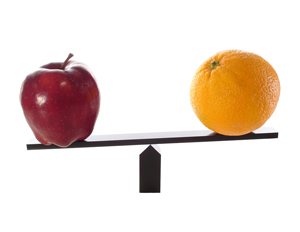
Comparing Apples to Apples
 Now, let’s go back to the issue I mentioned above–that ORAC values are normally calculated on the basis of 100-gram portions. The reason is that ORAC was originally developed to give data on whole foods, and 100 grams works out to just under a 4-ounce portion. It is essential, therefore, to make sure that the comparison cited for ORAC values is based on equivalent volumes (or servings). When sellers of mangosteen drinks claim ORAC values far superior to other antioxidants, are they comparing serving to serving? Probably not. In many cases, they have extended the numbers out to give the ORAC values in a liter/quart of mangosteen juice and then compared that to 1-ounce servings of other liquid antioxidant supplements. To get the true value per recommended 1-ounce serving, you would have to divide by 32, which takes you down to a more reasonable 500–600 ORAC units per serving. Don’t get me wrong: I like mangosteen and use it in some of my own formulations, but I don’t think it’s useful to exaggerate the numbers. And besides, as discussed above, since antioxidant levels in the blood are tightly regulated by your body, there are probably no health benefits to numbers over 3,000–5,000 ORAC per day of a single antioxidant anyway.
And when it comes to capsules, most capsules are 500 milligrams, which means it would take 56 capsules of a non-concentrated extract to equal an ounce of a food-based source of the antioxidants. In other words, it would take over 200 capsules to give you the same volume as a 4-ounce serving of the same antioxidant-rich whole food. If you want the same antioxidant potential in a single capsule, that means the ORAC value of the capsule needs to be 200 times more concentrated than the whole food in order to give you an equivalent value. This can be done by removing the water and fiber, which have no ORAC value. Grape skin extract, for example, has a much higher ORAC value than you’ll find in whole grape skins, but this does not mean that from a standpoint of cost, dose, and/or serving that the extract is necessarily superior. But keep in mind, there is the convenience factor. Isn’t it worth paying a premium to easily supplement with a full-spectrum antioxidant that works throughout the entire body and on all types of free radicals–an antioxidant that makes up for the fact that you aren’t including all necessary beneficial foods in your daily diet? When you can make it work, most definitely.
Antioxidant Foods
When it comes to antioxidants, your first choice should be to include antioxidant rich foods in your diet. It guarantees that you’re getting complete complexes and no synthetics. With that in mind, certain foods are high in antioxidants and should be a regular part of the diet. In fact, the U.S. Department of Agriculture recently rated a large number of foods according to their ORAC values. Remember, the higher the number, the more powerful the antioxidant effect. All ratings were based on 3.5 ounces of the tested food. As a reference, carrots (high in the carotenoids) had a 207 rating.
- Broccoli, 890
- Kale, 1,770
- Brussels sprouts, 980
- Blueberries, 2,400
- Spinach, 1,260
- Raisins, 2,830
- Strawberries, 1,540
- Prunes, 5,770
Don’t forget that these foods contain other phytonutrients that go well beyond their antioxidant value. Do not eat according to ORAC values alone. And do not think that you’ve covered your bases by focusing on looking for the one food with the highest antioxidant value. Incidentally, freeze dried acai berry probably tops the list at 104,000, but that’s not necessarily as good as it sounds because it’s very low in anthocyanins and phenolics. Remember, each high antioxidant food works in different areas of the body and on different free radicals. Eat as wide a variety of antioxidant rich foods as you can, as often as you can.
The Ultimate Antioxidant
Unfortunately, relying on foods alone for your antioxidants–although it’s extremely healthy–probably is not going to maximize the potential health benefits of antioxidants. As we have already discussed, it is virtually impossible to get all the antioxidants you need from diet alone–you would simply have to eat too much food to cover the full-spectrum of potential antioxidant benefits. And for reasons already discussed, you can’t get there by eating a single high antioxidant food as pushed by many companies. Yes, the ORAC value can be high, but no single food covers the full spectrum of your needs. And that is why you want to consider using a full spectrum antioxidant supplement. That said, there is really no such thing as an “ultimate” antioxidant supplement, but there are some that come close. You need to look for a formula that incorporates something approximating the scope listed below and that approximates the levels of each ingredient as listed. It may contain more of any particular ingredient, but if it contains substantially less, it’s merely there as label dressing (or “pixie dust” as we say in the trade). What follows is my current formulation that comes as close to “ultimate” as the real world allows, which is not to say that there aren’t other good formulas out there–just that I know this one. But again, I was guided by the things we’ve talked about so far:
- Get as wide a spectrum of antioxidants as possible so that they work in every area and every cell of your body.
- Use antioxidants that complement each other rather than duplicate each other.
- Use full complexes where feasible.
- Opt for natural over synthetic.
- Avoid all pixie dust ingredients. If an antioxidant can’t fit in the formula because it requires too many milligrams, don’t use it–don’t put it in simply to look good on the label.
With that in mind, here is the formula.
Natural Carotenoid Complex (5250 IU):
 Carotenoids are phytonutrients that protect plants from damage caused by UV radiation and other environmental factors. In humans, they have been shown to inhibit the proliferation of various types of cancer cells such as those affecting the lungs, stomach, cervix, breast, bladder, and mouth. They also have been proven to protect against atherosclerosis, cataracts, macular degeneration and other major degenerative disorders. The key carotenoids are: beta carotene, alpha carotene, lycopene, and zeaxanthin. The particular complex I selected for this formula contains several carotenoids, but emphasizes natural beta and alpha carotene.
Beta Carotene
Probably the best known of the carotenoids, beta carotene is converted by the body into vitamin A as needed to strengthen the immune system and promote healthy cell growth. In addition, beta carotene is a potent antioxidant, offering particular benefits to the immune system and the lungs.8 Hughes DA, Wright AJ, Finglas PM, et al. “The effect of beta-carotene supplementation on the immune function of blood monocytes from healthy male nonsmokers.” J Lab Clin Med. 1997 Mar;129(3):309-17. http://www.ncbi.nlm.nih.gov/pubmed/9042816 Note: synthetic beta carotene, which is made from acetylene gas, is to be avoided at all costs.9 Vitamin-Mineral Manufacturing Guide: Nutrient Empowerment, Vol. 1.+ Lakeport, CA: Nutrition Resource, 1986. Although “chemically” identical to natural beta carotene, synthetic beta carotene is structurally different and behaves very differently in the body. As a side note, synthetic beta carotene was used in all of the studies that produced negative results when beta carotene was tested. Also, it is important to understand that beta carotene is not the most important of the carotenoids. It’s just the only one that is a recommended daily requirement. Using a synthetic beta carotene isolate in supplements–or in studies–totally misses the mark. Once again, the FDA, the USDA, and most researchers are several decades behind the times.
Alpha Carotene
Recent studies have shown that alpha carotene is one of the most powerful carotenoids and has a strong inhibitory effect on the proliferation of various types of cancer cells such as those affecting the lungs,10 Min KB, Min JY. “Serum carotenoid levels and risk of lung cancer death in US adults.” Cancer Sci. 2014 Jun;105(6):736-43. http://www.ncbi.nlm.nih.gov/pubmed/24673770 stomach,11 Pelucchi C, Tramacere I, Bertuccio P, et al. “Dietary intake of selected micronutrients and gastric cancer risk: an Italian case-control study.” Ann Oncol. 2009 Jan;20(1):160-5. http://www.ncbi.nlm.nih.gov/pubmed/18669867 prostate,12 Umesawa M, Iso H, Mikami K, et al. “Relationship between vegetable and carotene intake and risk of prostate cancer: the JACC study.” Br J Cancer. 2014 Feb 4;110(3):792-6. http://www.ncbi.nlm.nih.gov/pubmed/24169341 breast,13 Pantavos A, Ruiter R, Feskens EF, et al. “Total dietary antioxidant capacity, individual antioxidant intake and breast cancer risk: The Rotterdam study.” Int J Cancer. 2014 Oct 4. http://www.ncbi.nlm.nih.gov/pubmed/25284450 bladder,14 Hung RJ, Zhang ZF, Rao JY, et al. “Protective effects of plasma carotenoids on the risk of bladder cancer.” J Urol. 2006 Sep;176(3):1192-7. http://www.ncbi.nlm.nih.gov/pubmed/16890724 and ovaries.15 Zhang M, Holman CD, Binns CW. “Intake of specific carotenoids and the risk of epithelial ovarian cancer.” Br J Nutr. 2007 Jul;98(1):187-93. http://www.ncbi.nlm.nih.gov/pubmed/17367574 It works by allowing normal cells to send growth-regulating signals to premalignant cells. Alpha carotene is usually found as part of a natural beta carotene complex, but is never found in synthetic isolates. In addition, studies have shown that people with the highest blood levels of vitamin D, a-carotene, ß-carotene, cryptoxanthin, lutein, and lycopene have a significantly reduced risk for melanoma.16 Millen AE, Tucker MA, et al. “Diet and melanoma in a case-control study.” Cancer Epidemiol Biomarkers Prev. 2004 Jun;13(6):1042-51. http://www.ncbi.nlm.nih.gov/pubmed/15184262
N-Acetyl-Cysteine (NAC) (225 mg)
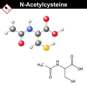 One of the keys to a healthy immune system is maintaining high levels of glutathione in the body. Unfortunately, supplementing with glutathione doesn’t really help. Fortunately, there are alternatives. Supplementation with N-acetyl-cysteine (NAC) has been proven to substantially raise the body’s glutathione levels.17 Asha Badaloo, Marvin Reid, Terrence Forrester, et al. “Cysteine supplementation improves the erythrocyte glutathione synthesis rate in children with severe edematous malnutrition1–3.” Am J Clin Nutr 2002;76:646–52. http://ajcn.nutrition.org/content/76/3/646.full.pdf+html In addition, NAC supplementation is mandatory for all smokers and big-city dwellers as it protects against toxic aldehydes that enter the body through cigarette smoke and pollution.18 Giorgia Volpi, Fabrizio Facchinetti, et al. “Cigarette smoke and a,ß-unsaturated aldehydes elicit VEGF release through the p38 MAPK pathway in human airway smooth muscle cells and lung fibroblasts.” Br J Pharmacol. Jun 2011; 163(3): 649–661. http://www.ncbi.nlm.nih.gov/pmc/articles/PMC3101625/ , 19 Carlsten C, MacNutt MJ, et al. “Anti-oxidant N-acetylcysteine diminishes diesel exhaust-induced increased airway responsiveness in person with airway hyper-reactivity.” Toxicol Sci. 2014 Jun;139(2):479-87. http://www.ncbi.nlm.nih.gov/pubmed/24814479
Another key benefit provided by NAC is its ability to modulate nitric oxide synthase expression and NF-kappaB activity, thus helping control inflammation throughout the body–e.g., in lung tissue,20 Blesa S, Cortijo J, Mata M, et al. “Oral N-acetyl cysteine attenuates the rat pulmonary inflammatory response to antigen.” Eur Respir J. 2003 Mar;21(3):394-400. http://erj.ersjournals.com/content/21/3/394.long liver tissue,21 Majano PL, Medina J, Zubia I, et al. “N-Acetyl-cysteine modulates inducible nitric oxide synthase gene expression in human hepatocytes.” J Hepatol. 2004 Apr;40(4):632-7. http://www.ncbi.nlm.nih.gov/pubmed/15030979
, 22 A El Hafiza, L El Wakeelb, et al. “High dose N-acetyl cysteine improves inflammatory response and outcome in patients with COPD exacerbations.” Egyptian Journal of Chest Diseases and Tuberculosis. Volume 62, Issue 1, January 2013, Pages 51–57. http://www.sciencedirect.com/science/article/pii/S0422763813000253 and intestinal tissue.23 Siddiqui A, Ancha H, Tedesco D, et al. “Antioxidant therapy with N-acetyl cysteine plus mesalamine accelerates mucosal healing in a rodent model of colitis.” Dig Dis Sci. 2006 Apr;51(4):698-705. http://www.ncbi.nlm.nih.gov/pubmed/16614991 Notably, this extends to NAC’s ability to both inhibit replication of the seasonal flu virus as well as inhibiting the expression of the pro-inflammatory cytokines triggered by the Type A flu virus.24 Geiler J, Michaelis M, et al. “N-acetyl-L-cysteine (NAC) inhibits virus replication and expression of pro-inflammatory molecules in A549 cells infected with highly pathogenic H5N1 influenza A virus.” Biochem Pharmacol. Feb 1;79(3):413-20. http://www.sciencedirect.com/science/article/pii/S000629520900728X As just mentioned, NAC has the ability to modulate NF-kappaB activity, and NF-kB is the “master signaling molecule” that regulates the activation of multiple inflammatory mediators.25 Kim H, Seo JY, Roh KH, et al. “Suppression of NF-kappaB activation and cytokine production by N-acetyl cysteine in pancreatic acinar cells.” Free Radic Biol Med. 2000 Oct 1;29(7):674-83. http://www.ncbi.nlm.nih.gov/pmc/articles/PMC3202196/pdf/10981515.pdf , 26 Chen G, Shi J, Hu Z, Hang C. “Inhibitory effect on cerebral inflammatory response following traumatic brain injury in rats: a potential neuroprotective mechanism of N-acetyl cysteine.” Mediators Inflamm. 2008;2008:716458. http://www.ncbi.nlm.nih.gov/pmc/articles/PMC2375967/
L-Methionine (225 mg)
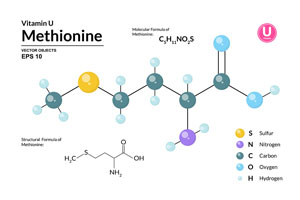 Methionine is an essential sulfur amino acid that works as a powerful antioxidant and liver detoxifier — where it assists in the normal detoxification processes. As an antioxidant, it provides powerful protection in the colon. Methionine is also involved in the synthesis of choline, adrenaline, lecithin, SAMe, and B12. The capacity of the body to excrete toxic substances through your urine is affected by the amount of L-methionine in the body. A deficiency can lead to fluid retention and edema. L-methionine is also responsible for the reduction of the level of histamine in the blood, which is why it can positively affect the symptoms of allergies. In addition, methionine plays a key role in helping to keep your body’s pH in balance as well as providing sulfur atoms for various chemical processes. And finally, L-methionine helps acidify your urine, which is important in preventing urinary tract infections, keeping in mind that E. coli, which is usually responsible for cystitis, cannot survive in an acidic environment.27 Günther M, Noll F, et al. “Harnwegsinfektprophylaxe — Urinansäuerung mittels L-Methionin bei neurogener Blasenfunktionsstörung.” Der Urologe (B). 2002; 218-220. http://link.springer.com/article/10.1007%2Fs00131-002-0207-x#page-1
Quercetin (180 mg)
 One of the class of antioxidants known as bioflavonoids, quercetin is a natural polyphenol flavonoid and is known to elicit both anti-inflammatory and antioxidant activities.28 Le NH, Kim CS, Park T, et al. “Quercetin Protects against Obesity-Induced Skeletal Muscle Inflammation and Atrophy.” Mediators Inflamm. 2014;2014:834294. http://www.ncbi.nlm.nih.gov/pubmed/25614714 It plays a primary role in preventing the release of histamines into the bloodstream as well as inhibiting inflammatory receptors and their signaling pathways (i.e., COX inhibition and TNF-a reduction), thereby helping to control food and pollen allergies as well as asthma.29 RT Ferreira, MAS Coutinho, et al. “Mechanisms Underlying the Antinociceptive, Antiedematogenic, and Anti-Inflammatory Activity of the Main Flavonoid from Kalanchoe pinnata.” Evid Based Complement Alternat Med. 2014; 2014: 429256. http://www.ncbi.nlm.nih.gov/pmc/articles/PMC4279175/ , 30 Townsend EA, Emala CW Sr. “Quercetin acutely relaxes airway smooth muscle and potentiates ß-agonist-induced relaxation via dual phosphodiesterase inhibition of PLCß and PDE4.” Am J Physiol Lung Cell Mol Physiol. 2013 Sep;305(5):L396-403. http://www.ncbi.nlm.nih.gov/pmc/articles/PMC3763034/ As an antioxidant, it protects the integrity of cell walls from free radical damage–even radiation induced free radical damage.31 Zbikowska HM, Antosik A, et al. “Does quercetin protect human red blood cell membranes against irradiation” Redox Rep. 2014 Mar;19(2):65-71. http://www.ncbi.nlm.nih.gov/pubmed/24257622 At low concentrations it can stimulate the “beneficial” proliferation of human cells,32 van der Woude H, Ter Veld MG, et al. “The stimulation of cell proliferation by quercetin is mediated by the estrogen receptor.” Mol Nutr Food Res. 2005 Aug;49(8):763-71. http://www.ncbi.nlm.nih.gov/pubmed/15937998 so it can be a potential drug in the treatment of neurodegenerative diseases; and, in high concentrations, it induces apoptosis thereby eliminating infected or abnormal cells and can serve as a potential anticancer drug with wide clinical application.33 Kobylinska A, Janas KM. “Health – promoting effect of quercetin in human diet.” Postepy Hig Med Dosw (Online). 2015 Jan 9;69(0):51-62. http://www.ncbi.nlm.nih.gov/pubmed/25589713
Ginkgo Biloba (180 mg, 24 percent ginkgo flavone glycosides and 6 percent terpene lactones)
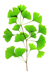 Known as the brain antioxidant, ginkgo improves blood flow to the brain34 Ameneh Mashayekh, Dzung L. Pham, et al. “Effects of Ginkgo biloba on cerebral blood flow assessed by quantitative MR perfusion imaging: a pilot study.” Neuroradiology. Mar 2011; 53(3): 185–191. http://www.ncbi.nlm.nih.gov/pmc/articles/PMC3163160/ (as well as to all the extremities) and acts as an antioxidant, thereby increasing brain functionality and helping to improve concentration and memory. The Ginkgo Evaluation of Memory (GEM) study published in 2012 seemed to close the door on Ginkgo when it concluded that Ginkgo does not lessen cognitive decline in older adults.35 Beth E. Snitz, Ellen S. O’Meara, et al. “Ginkgo biloba for Preventing Cognitive Decline in Older Adults.” JAMA. 2009;302(24):2663-2670. http://jama.jamanetwork.com/article.aspx?articleid=185120 On the other hand, a meta-analysis of seven other studies did indeed find benefits when using Ginkgo for Alzheimer’s.36 “Alzheimer’s disease: Can Ginkgo products help?” PubMed Health Last Update: July 18, 2013. http://www.ncbi.nlm.nih.gov/pubmedhealth/PMH0005095/ The analysis showed that people who took Ginkgo extract at double the dosage used in the GEM study were better able to perform daily activities again, like doing household chores or washing themselves. In line with the GEM study, the lower dose of Ginkgo extract (120 mg per day) detailed in the meta-study did not clearly influence the symptoms of Alzheimer’s disease. Also, Ginkgo extract may have a stronger benefit in younger patients or those who have psychological problems caused by Alzheimer’s disease. The trials also provided evidence that, when taken in high doses, Ginkgo could reduce psychological symptoms and improve people’s ability to remember things. It also seemed to reduce emotional stress for caregiving family members. When used for cognitive decline, it appears dosage matters. (Researchers do seem to love testing natural ingredients at low doses and then conclude they are ineffective.) In addition, Ginkgo’s anti-inflammatory, lung-relaxant properties have proven useful in the treatment of asthma, where it eases coughing and reduces tissue inflammation.37 Li M, Yange B, Yu H, Zhang H. “Clinical observation of the therapeutic effect of ginkgo leaf concentrated oral liquor on bronchial asthma.” Chinese Journal of Integrative Medicine. 1997;3:264–7. http://link.springer.com/article/10.1007%2FBF02934827#page-1 ,38 Chu X, Ci X, He J, et al. “A novel anti-inflammatory role for ginkgolide B in asthma via inhibition of the ERK/MAPK signaling pathway.” Molecules. 2011 Sep 6;16(9):7634-48. http://www.mdpi.com/1420-3049/16/9/7634 And finally, as a result of its ability to improve circulation in the extremities, there’s good evidence that ginkgo might ease leg pain caused by intermittent claudication, or clogging of the arteries in the legs.39 Max H Pittler, Edzard Ernst. “Ginkgo Biloba extract for the treatment of intermittent claudication: a meta-analysis of randomized trials.” The American Journal of Medicine. Volume 108, Issue 4, March 2000, Pages 276–281. http://www.sciencedirect.com/science/article/pii/S0002934399004544
Resveratrol (150 mg, 50 percent stabilized resveratrol)
 For several years, grape seed extract was all the rage; but as it turns out, grape “skin” was being mistakenly left out of the equation. It was subsequently discovered that grape skin contains its own powerful phytoalexin antioxidant called resveratrol. Thanks to early research, mostly done in test tubes and on animals, resveratrol gained renown as the secret ingredient in wine responsible for the French Paradox–that people who drank red wine were unaffected by high fat/high cholesterol diets. In truth, there is actually very little resveratrol in wine, and grapes are no longer used as the source for resveratrol extract. It turns out that levels in Japanese and Chinese knotweed are much higher than those found in grapes. So what are the benefits associated with resveratrol? In controlled studies, resveratrol has indeed been shown to reduce skin-cancer tumors by up to 98% and to stop production of leukemia cells.40 Moon, S.O., W. Kim, et al. “Resveratrol Suppresses Tumor Necrosis Factor-alpha-Induced Fractalkine Expression in Endothelial Cells.” Mol Pharmacol 70 (2006): 112–119. http://molpharm.aspetjournals.org/content/70/1/112.long Resveratrol also appears to have the ability to inhibit fatty acid signaling pathways and therefore may be helpful in the treatment of breast diseases.41 Khan A, Aljarbou AN, et al. “Resveratrol suppresses the proliferation of breast cancer cells by inhibiting fatty acid synthase signaling pathway.” Cancer Epidemiol. 2014 Dec;38(6):765-72. http://www.ncbi.nlm.nih.gov/pubmed/25448084 Studies also indicate that resveratrol can profoundly inhibit glucose uptake in HL-60 and U937 cells.42 Park JB. “Inhibition of glucose and dehydroascorbic acid uptakes by resveratrol in human transformed myelocytic cells.” J Nat Prod. 2001 Mar;64(3):381-4. http://www.ncbi.nlm.nih.gov/pubmed/11277764 Thus, resveratrol may prevent or abate metabolic disorders such as obesity and non-insulin dependent type 2 diabetes.43 Fiori JL, Shin YK, et al. “Resveratrol Prevents ß–cell Dedifferentiation in Non-Human Primates Given a High Fat/High Sugar Diet.” Diabetes. 2013 Oct;62(10):3500-13. http://www.ncbi.nlm.nih.gov/pmc/articles/PMC3781448/ In addition, it works as a cyclooxygenase (COX) inhibitor, thus halting the spread of cancer throughout the body. And although recent studies are ambivalent,44 Ponzo V, Soldati L, Bo S. “Resveratrol: a supplementation for men or for mice?” J Transl Med. 2014 Jun 3;12:158. http://www.ncbi.nlm.nih.gov/pmc/articles/PMC4049475/ some animal studies indicate it may slow down the aging process and prolong life.45 Baur, J.A., K.J. Pearson, et al. “Resveratrol Improves Health and Survival of Mice on a High-calorie Diet.” Nature 444 (November 2006): 337–342. http://www.ncbi.nlm.nih.gov/pubmed/17086191
Curcumin (120 mg)
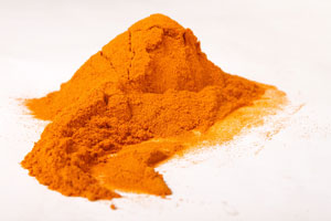 Curcumin is what gives turmeric its yellow color. Traditionally known for its anti-inflammatory effects, curcumin has been shown in the last 30 years to be a potent immunomodulatory agent that can modulate the activation of T cells, B cells, macrophages, neutrophils, natural killer cells, and dendritic cells.46 Jagetia GC, Aggarwal BB. “”Spicing up” of the immune system by curcumin.” J Clin Immunol. 2007;27(1):19–35. https://pubmed.ncbi.nlm.nih.gov/17211725-spicing-up-of-the-immune-system-by-curcumin/ It has also been shown that it can inhibit colon cancer cells by some 96% in a matter of hours.47 Vesna Milacic,Sanjeev Banerjee,et al. “Curcumin inhibits the proteasome activity in human colon cancer cells in vitro and in vivo.” Cancer Res. 2008 Sep 15; 68(18): 7283–7292. http://www.ncbi.nlm.nih.gov/pmc/articles/PMC2556983/ It also appears to have great potential in countering the effects of prostate cancer48 Marie-Hélène Teiten, François Gaascht, et al. “Chemopreventive potential of curcumin in prostate cancer.” Genes Nutr. Mar 2010; 5(1): 61–74. http://www.ncbi.nlm.nih.gov/pmc/articles/PMC2820199/ and breast cancer.49 Dongwu Liu and Zhiwei Chen. “The Effect of Curcumin on Breast Cancer Cells.” J Breast Cancer. Jun 2013; 16(2): 133–137. http://www.ncbi.nlm.nih.gov/pmc/articles/PMC3706856/ In a sense, curcumin can be thought of as natural chemotherapy–with the ability to selectively kill cancer cells, while at the same time leaving normal cells alone. Studies have also shown that curcumin can reduce the oxidative stress from diabetes by reducing the level of advanced glycation end products and the cross-linking of collagen.50 Stuart P. Weisberg, Rudolph Leibel, and Drew V. Tortoriello. “Dietary Curcumin Significantly Improves Obesity-Associated Inflammation and Diabetes in Mouse Models of Diabesity.” Endocrinology. Jul 2008; 149(7): 3549–3558. http://www.ncbi.nlm.nih.gov/pmc/articles/PMC2453081/ , 51 Renu A Kowluru and Mamta Kanwar. “Effects of curcumin on retinal oxidative stress and inflammation in diabetes.” Nutr Metab (Lond). 2007; 4: 8. http://www.ncbi.nlm.nih.gov/pmc/articles/PMC1868028/
Note: Curcumin and green tea strongly reinforce each other. Also, there is one caveat when supplementing with curcumin. A number of trials studying its efficacy and safety have demonstrated poor absorption and low bioavailability for most curcumin supplements, primarily because of its poor solubility in water. However, studies have shown that taking BioPerine (a black pepper extract) along with curcumin, significantly enhances both the serum concentration and the circulating bioavailability of curcumin.52 Preetha Anand, Ajaikumar B. Kunnumakkara, et al. “Bioavailability of Curcumin: Problems and Promises.” Mol. Pharmaceutics, 2007, 4 (6), 807-818. http://pubs.acs.org/doi/full/10.1021/mp700113r (More on BioPerine below.)
Green tea extract (120 mg, 95 percent polyphenols)
 Green tea antioxidants are of the same family as grape seed and pine bark extracts. They are polyphenols, chief of which are the flavonoids called proanthocyanidins. In green tea, the main proanthocyanidins are the catechins, and the most powerful of the catechins is epigallocatechin gallate (EGCG), found in the highest concentration in green tea. It works to prevent tumors from developing the blood vessels they need to survive (anti-angiogenesis),53 Chi Chiu Wang, Hui Xu, et al. “Prodrug of green tea epigallocatechin-3-gallate (Pro-EGCG) as a potent anti-angiogenesis agent for endometriosis in mice.” Angiogenesis. 2013 Jan;16(1):59-69. http://www.ncbi.nlm.nih.gov/pubmed/22948799 and it has been shown to inhibit metastasis.54 Chang CW, Hsieh YH, Yang WE, et al. “Epigallocatechingallate inhibits migration of human uveal melanoma cells via downregulation of matrix metalloproteinase-2 activity and ERK1/2 pathway.” Biomed Res Int. 2014;2014:141582. http://www.ncbi.nlm.nih.gov/pmc/articles/PMC4145379/ It is the first known natural telomerase inhibitor, eliminating the “immortality” of cancer cells, which is what makes them so deadly.55 Imad Naasani, Fujiko Oh-hashi, et al. “Blocking Telomerase by Dietary Polyphenols Is a Major Mechanism for Limiting the Growth of Human Cancer Cells in Vitro and in Vivo1.” Cancer Res February 15, 2003 63; 824. http://cancerres.aacrjournals.org/content/63/4/824.full Green tea is particularly effective in destroying the causes of leukemia, prostate cancer, and breast cancer. It has also been shown to be effective in regulating blood sugar, reducing triglycerides, and in reversing the ravages of heart disease.56 Hsieh SR, Cheng WC, et al. “Molecular targets for anti-oxidative protection of green tea polyphenols against myocardial ischemic injury.” Biomedicine (Taipei). 2014;4:23. http://www.ncbi.nlm.nih.gov/pmc/articles/PMC4264984/ (Incidentally, the Japanese, who drink large amounts of green tea, have some of the lowest rates of cardiovascular disease in the world.) Green tea seems to almost totally prevent cancer from causing DNA damage in smokers–a possible explanation as to why the Japanese, who are among the world’s heaviest smokers, have such a low incidence of lung cancer.57 Hakim IA, Chow HH, Harris RB. “Green tea consumption is associated with decreased DNA damage among GSTM1-positive smokers regardless of their hOGG1 genotype.” J Nutr. 2008 Aug;138(8):1567S-1571S. http://www.ncbi.nlm.nih.gov/pubmed/18641208 Finally, green tea has great benefits for the brain as well, serving as an effective monoamine oxidase (MAO) inhibitor, protecting against brain-cell death. The net result is that there are strong indications that green tea extract may play a major role in protecting against both Parkinson’s and Alzheimer’s disease.58 Ayokunle O. Ademosun and Ganiyu Oboh. “Comparison of the Inhibition of Monoamine Oxidase and Butyrylcholinesterase Activities by Infusions from Green Tea and Some Citrus Peels.” International Journal of Alzheimer’s Disease. Volume 2014 (2014), Article ID 586407, 5 pages. http://www.hindawi.com/journals/ijad/2014/586407/
Note: the consumption of casein from dairy products can completely block the absorption of the main catechins found in green tea.59 Egert S, Tereszczuk J, Wein S, et al. “Simultaneous ingestion of dietary proteins reduces the bioavailability of galloylated catechins from green tea in humans.” Eur J Nutr. 2013 Feb;52(1):281-8. http://www.ncbi.nlm.nih.gov/pubmed/22366739 In other words, drink your tea without milk, and take your green tea supplements separate from any dairy in your diet. Or, even better, just think of this as another reason to eliminate dairy from your diet.
Bilberry (120 mg, 25 percent anthocyanins)
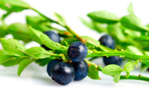 Although closely related to blueberries, bilberries are a distinct species. The anthocyanosides found in bilberries are known for their ability to help nourish and repair the tiny capillaries within the eye.60 Chung HK, Choi SM, Ahn BO, et al. “Efficacy of troxerutin on streptozotocin-induced rat model in the early stage of diabetic retinopathy.” Arzneimittelforschung. 2005;55(10):573-80. http://www.ncbi.nlm.nih.gov/pubmed/16294503 And although the stories about RAF pilots eating bilberries to improve night vision during WWII bombing missions is probably apocryphal, studies have indeed found that the antioxidants in bilberries can help with diseases of the eye such as macular degeneration.61 Fursova AZh, Gesarevich OG, et al. “[Dietary supplementation with bilberry extract prevents macular degeneration and cataracts in senesce-accelerated OXYS rats.]” Adv Gerontol. 2005;16:76-9. http://www.ncbi.nlm.nih.gov/pubmed/16075680 In addition, the bilberry bioflavonoids are beneficial to the connective tissue that both lines blood vessels and binds ligaments and which is often damaged or destroyed by systemic inflammation such as that seen in both arthritis and cardiovascular disease. 62 Karlsen A, Paur I, Bøhn S.V, et al. “Bilberry juice modulates plasma concentration of NF-?B related inflammatory markers in subjects at increased risk of CVD.” Eur J Nutr. 2010 2010 February 2;49:345–55. http://www.ncbi.nlm.nih.gov/pubmed/20119859 And one other nice benefit is that bilberry extract ameliorates hyperglycemia and insulin sensitivity through activation of the AMP-activated protein, kinase. Finally, in studies, bilberry extract has been shown to significantly reduce blood glucose concentrations and enhance insulin sensitivity.63 Takikawa M, Inoue S, Horio F, Tsuda T. “Dietary anthocyanin-rich bilberry extract ameliorates hyperglycemia and insulin sensitivity via activation of AMP-activated protein kinase in diabetic mice.” J Nutr. 2010 Mar;140(3):527-33. http://jn.nutrition.org/content/140/3/527.long
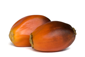 Tocotrienols (100 mg)
Derived from rice bran or palm oil, tocotrienols are a unique vitamin E fraction that is 40 times more powerful than standard vitamin E. The gamma-tocotrienol fraction, in particular, strongly inhibits both estrogen-responsive and the non-estrogen-responsive breast cancer cells.64 Kalanithi Nesaretnam, Puvaneswari Meganathan, et al. “Tocotrienols and breast cancer: the evidence to date.” Genes Nutr. Jan 2012; 7(1): 3–9. http://www.ncbi.nlm.nih.gov/pmc/articles/PMC3250526/ Over the last 30 years, a number of studies have shown that, thanks to their chemical structure, all four of the tocotrienols are more potent antioxidants than alpha-tocopherol65 Serbinova E, Kagan V, Han D, Packer L. “Free radical recycling and intramembrane mobility in the antioxidant properties of alpha-tocopherol and alpha-tocotrienol.” Free Radic Biol Med. 1991;10(5):263-75. http://www.ncbi.nlm.nih.gov/pubmed/1649783 and have a stronger anti-cancer effect.66 Constantinou C, Papas A, Constantinou AI. “Vitamin E and cancer: An insight into the anticancer activities of vitamin E isomers and analogs.” Int J Cancer. 2008 Aug 15;123(4):739-52. http://www.ncbi.nlm.nih.gov/pubmed/1649783 , 67 Wada S. “Chemoprevention of tocotrienols: the mechanism of antiproliferative effects.” Forum Nutr. 2009;61:204-16. http://www.ncbi.nlm.nih.gov/pubmed/19367124 The unsaturated side-chain in tocotrienols causes them to penetrate tissues with saturated fatty layers more efficiently.68 Suzuki YJ, Tsuchiya M, Wassall SR, et al. “Structural and dynamic membrane properties of alpha-tocopherol and alpha-tocotrienol: implication to the molecular mechanism of their antioxidant potency.” Biochemistry. 1993 Oct 12;32(40):10692-9.. http://www.ncbi.nlm.nih.gov/pubmed/8399214
Oligomeric Proanthocyanidins (100 mg, OPCs from grape seed extract) (95-97 percent polyphenols)
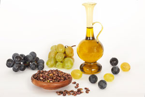 Similar to green tea, the active ingredients in grape seed extract are proanthocyanidins, although in a different combination and ratio (actually very similar to that found in pine bark extract). The importance of the proanthocyanidins in grape seed extract is that they are water-soluble and highly bioavailable. Grape seed extract is known as a defender of the circulatory system: it improves peripheral circulation, revives declining capillary activity by up to 140 percent, and increases vascular response by 82 percent.69 Godeau, R.L., C. Gavignet-Jeannin, et al. “The Effect of Procyanidolic Oligomers on Vascular Permeability: A Study Using Quantitative Morphology.” Pathol Biol (Paris) 38:6 (June 1990): 608–616. http://www.ncbi.nlm.nih.gov/pubmed/2165237 Studies have found that the OPCs in grape seed extract may be effective in relieving symptoms of chronic venous insufficiency (when veins have problems sending blood from the legs back to the heart) and reducing edema (swelling) after an injury or surgery.70 Belcaro G, Dugall M, et al. “Improvements of venous tone with pycnogenol in chronic venous insufficiency: an ex vivo study on venous segments.” Int J Angiol. 2014 Mar;23(1):47-52. http://www.ncbi.nlm.nih.gov/pubmed/24627617 , 71 Cesarone MR, Belcaro G, Rohdewald P, et al. “Improvement of signs and symptoms of chronic venous insufficiency and microangiopathy with Pycnogenol: a prospective, controlled study.” Phytomedicine. 2010 Sep;17(11):835-9. http://www.ncbi.nlm.nih.gov/pubmed/20579863 OPCs can repair varicose veins and aid in the prevention of bruising.72 Myburgh KH, Kruger MJ, Smith C. “Accelerated skeletal muscle recovery after in vivo polyphenol administration.” J Nutr Biochem. 2012 Sep;23(9):1072-9. http://www.ncbi.nlm.nih.gov/pubmed/22079208 And other studies have found beneficial effects for diabetic retinopathy.73 Li M, Ma YB, Gao HQ, et al. “A novel approach of proteomics to study the mechanism of action of grape seed proanthocyanidin extracts on diabetic retinopathy in rats.” Chin Med J (Engl). 2008 Dec 20;121(24):2544-52. http://www.ncbi.nlm.nih.gov/pubmed/?term=grape+seed+extract+retinopathy In addition, grape seed extract is synergistic with vitamin C, vastly increasing vitamin C activity and strengthening collagen activity, including in the connective tissue of the arterial wall and the skin. And OPCs have even shown benefit in treating breast cancer,74 Dinicola S, Pasqualato A, et al. “Grape seed extract suppresses MDA-MB231 breast cancer cell migration and invasion.” Eur J Nutr. 2014;53(2):421-31. http://www.ncbi.nlm.nih.gov/pubmed/23754570 prostate cancer,75 Kampa M, Theodoropoulou K, et al. “Novel oligomeric proanthocyanidin derivatives interact with membrane androgen sites and induce regression of hormone-independent prostate cancer.” J Pharmacol Exp Ther. 2011 Apr;337(1):24-32. http://www.ncbi.nlm.nih.gov/pubmed/21205921 and colon cancer.76 Kumar S, Kumar D, Raina K, et al. “Functional modification of adipocytes by grape seed extract impairs their pro-tumorigenic signaling on colon cancer stem cells and the daughter cancer cells.” Oncotarget. 2014 Oct 30;5(20):10151-69. http://www.ncbi.nlm.nih.gov/pmc/articles/PMC4259412/
Chaparral Extract (100 mg, 4:1 concentration)
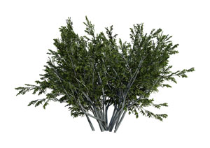 The active antioxidant in chaparral, a lignan called nordihydroguaiaretic acid (NDGA), has been shown to specifically target virtually all forms of herpes virus. Studies of a derivative of NDGA have proven to be even more effective than acyclovir, the prescription drug of choice, when it comes to treating the viruses HIV, HSV,77 Chen H, Teng L, Li JN, et al. “Antiviral activities of methylated nordihydroguaiaretic acids. 2. Targeting herpes simplex virus replication by the mutation insensitive transcription inhibitor tetra-O-methyl-NDGA.” J Med Chem. 1998 Jul 30;41(16):3001-7. http://www.ncbi.nlm.nih.gov/pubmed/9685239 and HPV78 Zhao J, Zhao Y, Chen W, et al. “The differentiation-inducing effect of Nordy on HPV-16 subgenes-immortalized human endocervical cells H8.” Anticancer Drugs. 2008 Aug;19(7):713-9. http://www.ncbi.nlm.nih.gov/pubmed/18594213 –HPV being notable as a primary factor in the onset of cervical cancer. Maybe even more important, there is no rapid buildup of drug resistance as happens with acyclovir.79 Chen, H., L. Teng, J.N. Li, et al. “Antiviral Activities of Methylated Nordihydroguaiaretic Acids.” J Med Chem 41:16 (July 1998): 3001–3007. http://www.ncbi.nlm.nih.gov/pubmed/9685239 And speaking of cervical cancer, studies have shown that NDGA and its derivatives are directly effective against cervical cancer itself.80 Gao P1, Zhai F, Guan L, Zheng J. “Nordihydroguaiaretic acid inhibits growth of cervical cancer SiHa cells by up-regulating p21.” Oncol Lett. 2011 Jan;2(1):123-128. http://www.ncbi.nlm.nih.gov/pmc/articles/PMC3412500/
According to a report in the May 2010 issue of the Medical Science Monitor, numerous studies have shown that NDGA is likely effective in the treatment of multiple diseases such as cardiovascular diseases, neurological disorders, and cancers, as well as being useful in the field of tissue engineering.81 Lü JM, Nurko J, et al. “Molecular mechanisms and clinical applications of nordihydroguaiaretic acid (NDGA) and its derivatives: an update.” Med Sci Monit. 2010 May;16(5):RA93-100. http://www.ncbi.nlm.nih.gov/pmc/articles/PMC2927326/ The report went on to explain that several medicinal properties such as NDGA’s antineoplastic, antiviral, and anti-inflammatory characteristics have been supported by in vitro and in vivo experimental studies, as well as historical reports. Studies have also confirmed that NDGA has extensive pharmacological effects and specific mechanisms of actions. It is a strong antioxidant; it can scavenge ROS (reactive oxygen species, another name for free radicals) or inhibit ROS production. It can stimulate nitrous oxide production, increase immune function, enhance central nervous system function, and prevent cardiovascular or other diseases. And tissue engineering studies demonstrate that NDGA-crosslinking is an effective way to improve the mechanical properties and biocompatibility of artificial tissues and organs. There are also strong indications that chaparral extract is an effective aid in the prevention of Alzheimer’s disease82 Goodman Y, Steiner MR, et al. “Nordihydroguaiaretic acid protects hippocampal neurons against amyloid beta-peptide toxicity, and attenuates free radical and calcium accumulation.” Brain Res. 1994;654:171–6. http://www.ncbi.nlm.nih.gov/pubmed/7982093 as well as rheumatoid arthritis.83 Eads D, Hansen R, Oyegunwa A, et al. “Terameprocol, a methylated derivative of nordihydroguaiaretic acid, inhibits production of prostaglandins and several key inflammatory cytokines and chemokines.” J Inflamm (Lond) 2009;6:2. http://www.ncbi.nlm.nih.gov/pmc/articles/PMC2631502/
Quite simply, when it comes to cancer, there are a number of studies that show the benefits of chaparral and NDGA–and many of them recent. For example, a 2010 study published in Bioorganic & Medicinal Chemistry Letters found that several synthetic variations of NDGA act as inhibitors against human liver cancer.84 Hwu JR, Hsu CI, et al. “Glycosylated nordihydroguaiaretic acids as anti-cancer agents.” Bioorg Med Chem Lett. 2011 Jan 1;21(1):380-2. http://www.ncbi.nlm.nih.gov/pubmed/21123067 The results of this study were reinforced by a 2014 study published in ChemMedChem journal that found that eight methylated versions of NDGA were protective against liver cancer.85 Hsu MH, Wu SC, et al. “Hepatocellular carcinoma targeting agents: conjugates of nitroimidazoles with trimethyl nordihydroguaiaretic Acid.” ChemMedChem. 2014 May;9(5):1030-7. http://www.ncbi.nlm.nih.gov/pubmed/24648164 And a 2012 study published in Breast Cancer Research and Treatment shows that it’s equally protective against breast cancer.86 Zhang Y, Xu S, Lin J, et al. “mTORC1 is a target of nordihydroguaiaretic acid to prevent breast tumor growth in vitro and in vivo.” Breast Cancer Res Treat. 2012 Nov;136(2):379-88. http://www.ncbi.nlm.nih.gov/pubmed/23053656 A 2008 study published in the journal Prostate concludes that it might be just as effective in dealing with prostate cancer.87 Ryan CJ, Zavodovskaya M, et al. “Inhibitory effects of nordihydroguaiaretic acid (NDGA) on the IGF-1 receptor and androgen dependent growth of LAPC-4 prostate cancer cells.” Prostate. 2008 Aug 1;68(11):1232-40. http://www.ncbi.nlm.nih.gov/pubmed/18491370 And finally, when it comes to cancer, chaparral is an effective counter to radiation-induced free-radical damage. (Note: if you’ve heard rumors about chaparral toxicity, it’s not what you’ve been told. Check out the section on chaparral in the report titled Cleansing Your Blood.)
Superoxide Dismutase (SOD) (75 mg, water-dispersible granules as opposed to oil based)
 The superoxide radical (O2-) is produced as a by-product of oxygen metabolism and causes many types of cell damage. Most notably, the superoxide radical attacks cell mitochondria.88 RAJINDAR S. SOHAL. “Mitochondria generate superoxide anion radicals and hydrogen peroxide.” The FASEB Journal. 1270 Vol. 11 December 1997. 1269-70. http://www.fasebj.org/content/11/14/1269.full.pdf When mitochondria are destroyed, the cell loses its ability to convert food to energy. It dies. In fact, superoxide may contribute to the pathogenesis (i.e., the development) of many diseases (the evidence is particularly strong for radiation poisoning and hyperoxic injury), and perhaps also to aging via the oxidative damage that it inflicts on cells.89 L. Benov. “How superoxide radical damages the cell.” 2001, Protoplasma Volume 217, Issue 1-3, pp 33-36. http://link.springer.com/article/10.1007/BF01289410 Superoxide dismutase (SOD) is an enzyme that facilitates the breakdown of the toxic superoxide radical into either ordinary molecular oxygen (O2) or hydrogen peroxide (H2O2). Hydrogen peroxide is also damaging, but less so than the superoxide radical and is degraded by catalase. (More on catalase later.) SOD works along with glutathione to neutralize reactive oxygen molecules in the body. SOD also works in the cytoplasm of the cell to prevent the hydroxyl radical from attacking enzymes, proteins, and the unsaturated fats in cell membranes. SOD is an extremely efficient enzyme; it catalyzes the neutralization of superoxide nearly as fast as the two can diffuse together spontaneously in solution.
But keep in mind, the key is not to eliminate all superoxide radicals, but to stop them from running wild. As we discussed earlier, superoxide plays a role both in controlling cancer and as a primary weapon of choice that your immune system uses to take on invading pathogens. To paraphrase Ecclesiastes 3:1, “To every superoxide radical there is a beneficial place in our body and a time when you will be thankful for its presence.” The problem is when they’re out of season, out of place, and out of control.
R-Lipoic Acid (50 mg)
Sometimes called the “Mother” antioxidant, alpha lipoic acid (ALA) plays a major role in helping recycle vitamins E and C so that they can be used over and over again by your body. Then, in its own right, ALA is one of the main boosters of glutathione levels in body cells,90 Busse E, Zimmer G, Schopohl B, Kornhuber B. “Influence of alpha-lipoic acid on intracellular glutathione in vitro and in vivo.” Arzneimittelforschung. 1992 Jun;42(6):829-31. http://www.ncbi.nlm.nih.gov/pubmed/1418040 and is one of the key co-factors involved both in generating energy in the cell’s mitochondria and in protecting those mitochondria from damage.91 Giorgia Mellia, Michela Taianaa, et al. “Alpha-lipoic acid prevents mitochondrial damage and neurotoxicity in experimental chemotherapy neuropathy.” Experimental Neurology. Volume 214, Issue 2, December 2008, Pages 276–284. http://www.sciencedirect.com/science/article/pii/S0014488608003312 This is extremely important as increasing evidence shows that mitochondrial dysfunction due to the oxidation of lipids, proteins, and nucleic acids plays an important role in brain aging and neurodegenerative diseases such as Alzheimer, Parkinson’s, ALS, and Huntington disease.92 Ashu Johri and M. Flint Beal. “Mitochondrial Dysfunction in Neurodegenerative Diseases.” J Pharmacol Exp Ther. 2012 Sep; 342(3): 619–630. http://www.ncbi.nlm.nih.gov/pmc/articles/PMC3422529/ , 93 Lin MT1, Beal MF. “Mitochondrial dysfunction and oxidative stress in neurodegenerative diseases.” Nature. 2006 Oct 19;443(7113):787-95. http://www.ncbi.nlm.nih.gov/pubmed/17051205 Mitochondria provide energy for basic metabolic processes, produce oxidants as inevitable by-products, and decay with age, impairing cellular metabolism and leading to cellular decline.94 Hardas SS, Sultana R, Clark AM, et al. “Oxidative modification of lipoic acid by HNE in Alzheimer disease brain.” Redox Biol. 2013 Jan 30;1:80-5. http://www.ncbi.nlm.nih.gov/pmc/articles/PMC3757677/ In addition to its involvement with mitochondria, various mechanisms for ALA’s positive effects on cognitive function have been suggested, including improvement of memory-related signaling pathways, reduction of oxidative stress, and restoring the activity of acetylcholinesterase and Na + , K + -ATPase. Aging reduces the activity of acetylcholinesterase in the cerebral cortex, cerebellum, striatum, hippocampus, and hypothalamus; supplementation with ALA reverses that decrease. Also, treatment with ALA protects cortical neurons against cytotoxicity induced by beta-amyloid or hydrogen peroxide.95 Lei Zhanga, Gou qaing Xingb, et al. “a-lipoic acid protects rat cortical neurons against cell death induced by amyloid and hydrogen peroxide through the Akt signalling pathway.” Neurosci Lett. 2001 Oct 26;312(3):125-8. http://www.ncbi.nlm.nih.gov/pubmed/11602326 
But it’s not just your brain. A review of several studies has shown that ALA may help relieve several risk factors associated with metabolic syndrome–a collection of symptoms that can lead to diabetes and heart disease. As the review explained, ALA can reduce blood pressure and insulin resistance, improve lipid profiles, and promote weight-loss.96 Pershadsingh HA. “Alpha-lipoic acid: physiologic mechanisms and indications for the treatment of metabolic syndrome.” Expert Opin Investig Drugs. 2007 Mar;16(3):291-302. http://www.ncbi.nlm.nih.gov/pubmed/17302524 In fact, studies have shown that ALA supplementation can even be beneficial in cases of full blown diabetes, actually resulting in an improvement in islet function.97 Zhao L1, Hu FX. “a-Lipoic acid treatment of aged type 2 diabetes mellitus complicated with acute cerebral infarction.” Eur Rev Med Pharmacol Sci. 2014 Dec;18(23):3715-9. http://www.europeanreview.org/article/8177
Alpha-lipoic acid exists in three different forms: alpha R-lipoic acid, alpha S-lipoic acid, and alpha RS-lipoic acid. Alpha S-lipoic acid and alpha RS-lipoic acid are synthetics; these are the forms commonly known and sold as alpha-lipoic acid supplements. On the other hand, alpha R-lipoic acid (better known as R-lipoic acid) is the only version of lipoic acid that exists in nature. It is synthesized by both plants and animals. Supplements sold as R-lipoic acid contain only the natural (cis) form of lipoic acid. They are twice as effective as the synthetic (trans) form supplements sold as alpha lipoic acid. Thus, formulas that use R-lipoic acid require only about 50 mg per serving.
Lutein (10 mg)
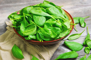 Lutein is a xanthophyll carotenoid and is synthesized only by plants. Like other xanthophylls, it is found in high quantities in green leafy vegetables such as spinach, kale, and yellow carrots. It can also be found in egg yolk and animal fats, provided the animals consumed lutein in their feed. In fact, lutein is now frequently used in chicken feed to improve the color of broiler chicken skin and to produce darker yellow egg yolk. In addition to being a specific for the prevention of macular degeneration (lowering the risk by over 50%),98 Richer S, Stiles W, et al. “Double-masked, placebo-controlled, randomized trial of lutein and antioxidant supplementation in the intervention of atrophic age-related macular degeneration: the Veterans LAST study (Lutein Antioxidant Supplementation Trial).” Optometry. 2004 Apr;75(4):216-30. http://www.ncbi.nlm.nih.gov/pubmed/15117055 lutein has also been shown to have strong anti-cancer properties.99 Slattery ML, Benson J, Curtin K, Ma KN, Schaeffer D, Potter JD. “Carotenoids and colon cancer.” Am J Clin Nutr 2000 Feb;71(2):575-82. http://ajcn.nutrition.org/content/71/2/575.long Its antioxidant effect is significantly enhanced by the presence of zeaxanthin and bilberry. There is also evidence that increasing lutein and zeaxanthin intake lowers the risk of cataract development.100 SanGiovanni JP, Chew EY, Clemons TE, et al. “The relationship of dietary carotenoid and vitamin A, E, and C intake with age-related macular degeneration in a case-control study: AREDS Report No. 22.” Arch Ophthalmol. 2007 Sep;125(9):1225-32. http://www.ncbi.nlm.nih.gov/pubmed/17846363 Lutein is one of two major carotenoids found as a color pigment in the human eye (macula and retina). It is thought to function as a light filter, protecting the eye tissues from sunlight damage.
Lycopene (6 mg)
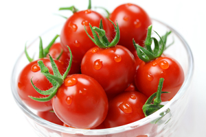 Lycopene is a bright red carotene pigment and is the phytochemical found in tomatoes and many other red fruits and vegetables, such as guavas, watermelon, and papayas–but not strawberries. An interesting note is that cooking actually enhances its bioavailability. Studies that looked at lycopene levels in the blood found that levels were higher after people ate cooked tomatoes than after they ate raw tomatoes or drank tomato juice. Lycopene appears to be one of the best defenses against prostate and bladder cancers.101 Hung RJ When used by itself, study results are mixed with newer studies less promising than older studies, but when used in conjunction with green tea extract, studies indicate that lycopene inhibits prostate cancer proliferation by 90 percent.102 Jian L, Lee AH, Binns CW. “Tea and lycopene protect against prostate cancer.” Asia Pac J Clin Nutr. 2007;16 Suppl 1:453-7. http://apjcn.nhri.org.tw/server/APJCN/16%20Suppl%201//453.pdf Now, it should be noted that one of the reasons that more recent studies with lycopene started producing less favorable results is that PSA screening started detecting more “cancers” that were missed in earlier studies, thus bringing down the “percentage” of favorable results. However, that data may be misleading as two-thirds of those reported cancers may have been false positives. The PSA test has recently been determined to be highly unreliable. In fact, even the discoverer of the PSA test now says that the test “is hardly more effective than a coin toss.” When you correct for that bad data, the numbers for lycopene start to become more favorable again.
BioPerine® (4500 mcg)
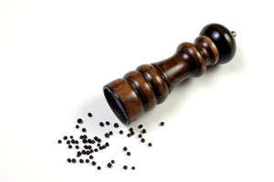 BioPerine is an extract obtained from black pepper fruit grown in southern India. It serves the same purpose in this formula that cayenne serves in liquid tinctures; it drives the other ingredients into the blood and significantly improves their efficacy. When BioPerine was administered with beta-carotene (which is fat soluble), the observed result was a 60% increase in blood level beta-carotene.103 Badmaev, V, Majeed, M. et al. “Piperine, An Alkaloid Derived from Black Pepper, Increases Serum Response of Beta-Carotene During 14 Days of Oral Beta-Carotene Supplementation.” Nutrition Research, 19(3) 381-388, 1999. http://www.sciencedirect.com/science/article/pii/S027153179900007X Similar results were obtained with vitamin B6 (which is water soluble) and the mineral selenium, suggesting that BioPerine significantly increases absorption whether the nutraceutical is fat or water soluble. Notably, in human studies, the bioavailability of curcumin was increased twentyfold when used with BioPerine.104 Shoba G, et al. “Influence of piperine on the pharmacokinetics of curcumin in animals and human volunteers.” Planta Med; 64(4):353-6. 1998. http://www.ncbi.nlm.nih.gov/pubmed/9619120 BioPerine improves the absorption of many vitamins and biochemicals in the intestine, promotes their retention in cells, and prevents their being metabolized in the liver. Effectively, it increases the efficacy of virtually all of the ingredients in this formula.
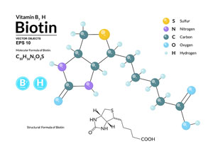 Biotin (1.8 mg)
The chemical structure of biotin is similar to that of alpha lipoic acid (ALA), and there is some evidence that high concentrations of ALA can compete with biotin for transport across cell membranes.105 Prasad PD, Wang H, Huang W, et al. “Molecular and functional characterization of the intestinal Na+-dependent multivitamin transporter.” Arch Biochem Biophys. 1999;366(1):95-106. http://www.ncbi.nlm.nih.gov/pubmed/10334869 , 106 Balamurugan K, Vaziri ND, Said HM. “Biotin uptake by human proximal tubular epithelial cells: cellular and molecular aspects.” Am J Physiol Renal Physiol. 2005;288(4):F823-831. http://ajprenal.physiology.org/content/288/4/F823 Further, the administration of high doses of ALA by injection to rats decreased the activity of two biotin-dependent enzymes by about 30%-35% (98).107 Zempleni J, Trusty TA, Mock DM. “Lipoic acid reduces the activities of biotin-dependent carboxylases in rat liver.” J Nutr. 1997;127(9):1776-1781. http://jn.nutrition.org/content/127/9/1776.long The bottom line is that when taking high doses of alpha lipoic acid, it is advisable to also take biotin to counter any tendency ALA may have to compete with biotin and interfere with its activity in the body.
Zeaxanthin (500 mcg)
Lutein and zeaxanthin are both part of a group of carotenoids known as xanthophylls, which are extremely beneficial to the eyes and help significantly in the prevention of macular degeneration,108 Liu R, Wang T, Zhang B, et al. “Lutein and zeaxanthin supplementation and association with visual function in age-related macular degeneration.” Invest Ophthalmol Vis Sci. 2014 Dec 16;56(1):252-8. http://www.ncbi.nlm.nih.gov/pubmed/25515572 In fact, zeaxanthin and lutein are the two primary xanthophyll carotenoids found in the retina of the eye. Within the central macula itself, zeaxanthin is the dominant carotenoid, whereas in the peripheral retina, lutein is dominant. And it’s not just in your eye, zeaxanthin is one of the most common carotenoids found in nature, and like lutein, it is found in the highest quantity in the leaves of green plants. But it is also found in green, yellow, and orange vegetables such as broccoli, green peas, Brussels sprouts, goji berries, and orange bell peppers. Also, like lutein, animals cannot produce it. They get what they need by eating sources rich in zeaxanthin. For this reason, it can also be found in animal fats and skin and in egg yolks.
We mentioned earlier how beta and alpha carotene, lutein, and lycopene lower the risk of melanoma. Zeaxanthin, however, plays a different role in preventing the same disease. As it turns out, zeaxanthin has a “selectively cytotoxic effect on melanoma cells,” actually inducing apoptosis in those cells.109 Ming-Chao Bi, Richard Rosen, et al. “Zeaxanthin Induces Apoptosis in Human Uveal Melanoma Cells through Bcl-2 Family Proteins and Intrinsic Apoptosis Pathway.” Evid Based Complement Alternat Med. 2013; 2013: 205082. http://www.ncbi.nlm.nih.gov/pmc/articles/PMC3810440/
Catalase (300 mcg)
 Glutathione peroxidase, superoxide dismutase, and catalase are the primary three enzymes produced in the body as an antioxidant defense. The first two we’ve already covered, so let’s take a look at catalase. Catalase is a specific for protection against tumors, and a little can go a long way. One catalase enzyme molecule can catalyze the breakdown of forty million molecules of peroxide radicals into water and oxygen in just one minute. In addition to acting as a super antioxidant, catalase also has the ability to use hydrogen peroxide to oxidize toxins including methanol, ethanol, formic acid, formaldehyde, and nitrite. This type of dual activity makes it a crucial cellular enzyme.
A 2005 study published in Science found that catalase plays a major role in controlling free radical damage and extending lifespan.110 Schriner SE, Linford NJ, Martin GM, et al. “Extension of murine life span by overexpression of catalase targeted to mitochondria.” Science. 2005 Jun 24;308(5730):1909-11. http://www.ncbi.nlm.nih.gov/pubmed/15879174 As the study’s authors concluded, “These results support the free radical theory of aging and reinforce the importance of mitochondria as a source of these radicals.” Meanwhile, a 2006 study published in the Journal of Cellular and Molecular Medicine found that oxidative stress, and its accumulation in protein as well as DNA damage are all reduced by the increased activity of catalase.111 Sinitsyna O, Krysanova Z, et al. “Age-associated changes in oxidative damage and the activity of antioxidant enzymes in rats with inherited overgeneration of free radicals.” J Cell Mol Med. 2006 Jan-Mar;10(1):206-15. http://www.ncbi.nlm.nih.gov/pubmed/16563232 This study also found that increased catalase activity leads to reduced mitochondrial dysfunction and slows the process of aging in tested animals.
Selenium (55 mcg, as methylselenocysteine)
 Selenium in both foods and in supplements comes in either an inorganic form (selenite or selenate) or in an organic form. In living systems, the organic form of selenium is found in the amino acids selenomethionine, selenocysteine, and methylselenocysteine. Selenium is actually transformed from inorganic to organic forms by lactic acid bacteria as found mostly on the skin of healthy fruits and vegetables.112 Sarang Dilip Pophalya, Poonama, et al. “Selenium enrichment of lactic acid bacteria and bifidobacteria: A functional food perspective.” Trends in Food Science & Technology. Volume 39, Issue 2, October 2014, Pages 135–145. http://www.sciencedirect.com/science/article/pii/S0924224414001587 The most useful of its organic forms is methylselenocysteine. The advantage of the organic form is that it offers improved absorbability while at the same time significantly lessening concerns about toxicity. How much less toxic is organic selenium? Studies have shown that inorganic selenium is toxic in the human body at levels as low as 1 ppm, whereas organic selenium has no toxic impact even at levels five times that.113 Nagy G, Benko I, Kiraly G, et al. “Cellular and nephrotoxicity of selenium species.” J Trace Elem Med Biol. 2015 Jan 6. pii: S0946-672X(14)00262-4. http://www.ncbi.nlm.nih.gov/pubmed/25604949
Once inside the body, selenium becomes a component of glutathione peroxidase, which is primarily responsible for reducing peroxide free radicals that include lipid peroxide formation in cell membranes (formed as fats are oxidized). Reduction of peroxides breaks the auto-oxidative chain reaction that damages cell membranes. Selenium is synergistic with glutathione and catalase in helping to protect the integrity of cell membranes. It also stops the growth of tumors, and it protects the liver. It’s also important for healthy thyroid and immune system function.
The recommended daily allowance for selenium (based on studies with inorganic selenium) is 55 micrograms. But as with most RDI’s, that’s too low. The reason it’s set so low is because of concerns about toxicity. The tolerable upper intake level for selenium is 400 micrograms a day for adults. At first this might sound high, but consider that the average American gets 80 to 110 micrograms of selenium a day from the foods they eat. Selenium is found in seafood, red meat, and from food grown in selenium-rich roil (and that, by the way, is a big wild card as most soil on well worked farmland is selenium deficient). Incidentally, one ounce of Brazil nuts “may” contain more than 500 micrograms of selenium. But what really compounds the problem is that when I designed my first full-spectrum antioxidant formula several decades ago, nobody was adding selenium to their supplements. I could maximize the amount of organic selenium with no concerns for toxicity. Now, almost every vitamin pill on the market contains added selenium–and in anything you buy outside the health food store it’s almost always the inexpensive, inorganic form of selenium. The Centrum formulas for both men and women, for example, each contains 55 mcg of inorganic selenium.114 http://www.centrum.ca/learn/vitamins-minerals/selenium Plus many of the foods you now eat are “fortified” with inorganic selenium. For that reason, I’ve cut the level of organic selenium I use in this formula to just 55 mcg–just enough to cover any dietary deficiencies, but low enough not to present a problem if you’re already getting multiple shots of selenium in your daily regimen. Incidentally, the reason you want to be sure you’re getting enough selenium is because low levels have been connected to death from everything ranging from heart disease to many different kinds of cancer.115 “Selenium.” NIH July 02, 2013. (Accessed 28 Jan 2015.) http://ods.od.nih.gov/factsheets/Selenium-HealthProfessional/#en36
Other Antioxidants
There are other antioxidant supplements that are also well worth taking. They aren’t included in the formula above for the simple reason that the quantity needed for these antioxidants to be effective is too large to be incorporated into a full-spectrum formula. They need to be taken separately or in larger capacity formulas. For example, I use a number of them in my superfood formula in which servings are measured out in large scoops rather than small capsules. Some of these supplements are:
- Red Raspberry (40 mg, active ellagitannins)–Scientific studies have proven that supplementation with 40 mg per day of red raspberry ellagitannins prevents the development of cancer cells.116 Ayrton, A.D., et al. “Antimutagenicity of Ellagic Acid Towards the Food Mutagen IQ: Investigation into Possible Mechanisms of Action.” Food Chem Toxicol 30:4 (1992): 289-95. http://www.ncbi.nlm.nih.gov/pubmed/1628864 , 117 Constantinou, A., et al. “The Dietary Anticancer Agent Ellagic Acid is a Potent Inhibitor of DNA Topoisomerases in Vitro.” Nutr Cancer 23:2 (1995):121–130. http://www.ncbi.nlm.nih.gov/pubmed/7644381 (Note: It takes a full cup of red raspberry puree or 1000 mg or more of a red raspberry seed powder to get 40 mg of active ellagitannins.) At low concentrations, ellagitannins slow the growth of cancer cells; at higher concentrations, they force cancer cells to self-destruct. There are number of supplements that sell pure isolated ellagic acid, but this form does not work as well as the full ellagitannin complex and may produce side effects such as high blood pressure.
 Pomegranate (500 mg ellagitannins)–In the past few years, studies of pomegranates have shown components that inhibit skin and breast cancers, high blood pressure, and atherosclerosis. Pomegranate juice is an antioxidant powerhouse, packed with both the polyphenols that make red wine and green tea effective and the ellagitannins that produce the health benefits of red raspberries. According to an article in the Journal of Clinical Nutrition, people who drank just 2 ounces of pomegranate juice a day for a week increased antioxidant activity by 9 percent.118 Aviram, M., L. Dornfeld, M. Rosenblat, et al. “Pomegranate Juice Consumption Reduces Oxidative Stress, Atherogenic Modifications to LDL, and Platelet Aggregation: Studies in Humans and in Atherosclerotic Apolipoprotein E-Deficient Mice.” Am J Clin Nutr 71:5 (2000): 1062–1076. http://www.ncbi.nlm.nih.gov/pubmed/10799367 Supplementation with pomegranate extract makes sense if you are looking at cancer prevention, maintaining a healthy heart, reducing LDL (“bad”) cholesterol, lowering blood pressure, and protecting against free radicals and aging. In fact, use both red raspberry and pomegranate: the mix of ellagitannins in the two foods appear to be both different and complementary.
- Noni Leaf (500 mg; or noni fruit extract concentrated 5:1)–The active biochemical in noni powder, proexeronine, is readily converted in the body to xeronine, an alkaloid that is necessary in virtually every cell in the body. Raising the levels of xeronine to optimum levels can produce profound health effects, and noni appears unique in its ability to accomplish this.119 Serafini MR1, Santos RC, et al. “Morinda citrifolia Linn leaf extract possesses antioxidant activities and reduces nociceptive behavior and leukocyte migration.” J Med Food. 2011 Oct;14(10):1159-66. http://www.ncbi.nlm.nih.gov/pubmed/21548805 It also triggers the release of nitric oxide from the body’s cells, a key component in optimizing blood pressure,120 Katherine C. Wood, Miriam M. Cortese-Krott, Jason C. Kovacic, et al. “Circulating Blood eNOS Contributes to the Regulation of Systemic Blood Pressure and Nitrite Homeostasis.” Arterioscler Thromb Vasc Biol. Aug 2013; 33(8). http://www.ncbi.nlm.nih.gov/pmc/articles/PMC3864011/ alleviating erectile dysfunction, and in fighting viral, bacterial, and parasitic infections, and the development of tumors. It is also associated with optimizing learning, memory, and sleep, and reducing the pain associated with inflammation and rheumatoid arthritis.121 Isla S Mackenzie, Daniel Rutherford,Thomas M MacDonald. “Nitric oxide and cardiovascular effects: new insights in the role of nitric oxide for the management of osteoarthritis.” Arthritis Res Ther. 2008; 10(Suppl 2): S3. http://www.ncbi.nlm.nih.gov/pmc/articles/PMC2582806/
- Goji (500 mg, 40 percent polysaccharides)–Studies have shown that goji berry can induce an immune response and may possess potential therapeutic efficacy in cancer.122 Gan, L., S.H. Zhang, Q. Liu, et al. “A Polysaccharide-Protein Complex from Lycium barbarum Upregulates Cytokine Expression in Human Peripheral Blood Mononuclear Cells.” Eur J Pharmacol 471:3 (June 2003): 217–222. http://www.ncbi.nlm.nih.gov/pubmed/12826241 Other studies have demonstrated that goji induces a remarkable adaptability to exercise load plus enhanced resistance and accelerated elimination of fatigue.123 Luo, Q., J. Yan, S. Zhang. “Isolation and Purification of Lycium barbarum Polysaccharides and Its Anti-fatigue Effect.” Wei Sheng Yan Jiu 29:2 (March 2000): 115–117. http://www.ncbi.nlm.nih.gov/pubmed/12725093 And finally, goji has demonstrated the ability to prevent and reverse the effects of free-radical damage.124 Zhang, X. “Experimental Research on the Role of Lycium barbarum Polysaccharide in Anti-peroxidation.” Zhongguo Zhong Yao Za Zhi 18:2 (February 1993): 110–112, 128. http://www.ncbi.nlm.nih.gov/pubmed/8323695
- Mangosteen (500 mg)–Mangosteen contains a unique group of antioxidants called xanthones. Xanthones, particularly beta and gamma mangostin, maintain the immune system, support cardiovascular health, optimize joint flexibility, are naturally antibiotic, antiviral, and anti-inflammatory, and are some of the most powerful antioxidants found in nature (over 14 times the ORAC value of red raspberries–but not ellagitannins).125 Fabiola Gutierrez-Orozco and Mark L. Failla1. “Biological Activities and Bioavailability of Mangosteen Xanthones: A Critical Review of the Current Evidence.” Nutrients. Aug 2013; 5(8): 3163–3183. http://www.ncbi.nlm.nih.gov/pmc/articles/PMC3775248/ In addition, recent studies have confirmed that gamma mangostin is a potent cyclooxygenase (COX) inhibitor, an important factor in reducing inflammation, pain, and fever. And a number of studies have demonstrated a pronounced ability to suppress tumor growth in breast cancer, prostate cancer, glioblastoma, and colon cancer patients.126 Gutierrez-Orozco
- L-Carnosine (500–1,500 mg)–L-carnosine is a naturally occurring combination of two amino acids, alanine and histidine that can actually reverse the signs of aging. It works as an antioxidant to protect cellular protein from attack by carbonyl groups and prevents the oxidation of sugars in the body. Supplementation with carnosine represents one of the most powerful things you can do to hold back the ravages of old age.
- Vitamin E (500 mg of mixed tocopherols and tocotrienols)–We have talked a good deal about the health benefits of vitamin E already. Just make sure you use a complete vitamin E complex that contains all four tocopherols and all four tocotrienols–or a pair of supplements that provide the full vitamin E complex. Don’t settle for just d-alpha, because, at best, it’s only the sixth most beneficial component–and it’s incomplete. And of course, dl-alpha should be a non-starter by this point.
- Vitamin C (500–1,000 mg)–The antioxidant benefits of vitamin C are invaluable. The trick is that almost all of the vitamin C sold today is in an isolate form that is extremely harsh on the body. Yes, I know that’s the form that Linus Pauling studied, but nobody’s perfect. You need 500–1,000 mg of vitamin C a day, but try to find it in a “living-food matrix,” that is, bound to food so your body can effectively utilize it.
Conclusion
Over the years, I have created a number of antioxidant formulas for supplement companies. In fact, this is my specialty–the design of cutting-edge nutritionals using standardized herbs and isolates. On the other hand, I have also become known as a strong advocate for the use of whole herbs and food complexes in all formulations. Why the apparent contradiction? As we’ve already discussed, nature packages nutrients in complexes and, in general, it is best to design supplements using only whole-food complexes. Antioxidants, however, provide one of the few exceptions to that rule, as long as a few provisos are followed.
The reason for the exception is that antioxidants are specialists, not generalists. No single antioxidant works on all free radicals and in every area of the body. For example, glutathione protects and repairs the liver, whereas bilberry works to defend the eyes. You need to combine a number of antioxidants in one supplement in order to offer an effective full-spectrum defense. In fact, many antioxidants reinforce and/or recycle each other.
For maximum protection, it is vital that you get an antioxidant complex that provides a full-spectrum defense. This is the only real defense against free-radical devastation. (Researchers who test single ingredient antioxidants are missing the point.) For this reason, it is impossible to even come close to fitting the variety you need (at adequate levels) into one supplement–unless you use some standardized herbs and isolates. The trick is to:
- Use only natural isolates, no synthetics.
- Get as complete a complex as you can, even when using isolates.
- Take advantage of the synergistic effect that a number of the antioxidants share with each other. For example, zeaxanthin and lutein reinforce each other, as do curcumin and green tea.
As we discussed earlier, it is virtually impossible to get all the antioxidants you need from diet alone–you would simply have to eat too much food to cover the full-spectrum of potential antioxidant benefits. The bottom line is that a good, antioxidant formula should play a key role in your health supplement regimen. Find a formula you like and use it daily. Remember, it is a “supplement” program–use it to supplement an antioxidant-rich diet, not to replace it.
(Originally published 03/01/2004. Updated 02/23/2015.)
References
| ↑1 | Stephen Barrett, M.D. “Antioxidants and Related Phytochemicals: Current Scientific Perspective.” Quackwatch November 14, 2012. (Accessed 28 Jan 2015.) http://www.quackwatch.org/03HealthPromotion/antioxidants.html |
|---|---|
| ↑2 | “UCL study questions basis for treatment of diseases including cancer and arthritis” UCL News 26 February 2004. (Accessed 29 Jan 2015.) http://www.ucl.ac.uk/news/news-articles/news-releases-archive/oxygen |
| ↑3 | de Rijk MC, Breteler MM, et al. “Dietary antioxidants and Parkinson disease. The Rotterdam Study.” Arch Neurol. 1997 Jun;54(6):762-5. http://www.ncbi.nlm.nih.gov/pubmed/9193212 |
| ↑4 | “Free Radical Cancer Biology Program.” University of Iowa Hospitals & Clinics. (Accessed 29 Jan 2015.) http://www.uihealthcare.org/otherservices.aspx?id=21027 |
| ↑5 | Etienne Pigeolet, Philippe Corbisier, et al. “Glutathione peroxidase, superoxide dismutase, and catalase inactivation by peroxides and oxygen derived free radicals.” Mech Ageing Dev. 1990 Feb 15;51(3):283-97. http://www.ncbi.nlm.nih.gov/pubmed/2308398 |
| ↑6 | B H Segal, P Veys, H Malech, M J Cowan. “Chronic Granulomatous Disease: Lessons from a Rare Disorder.” Biol Blood Marrow Transplant. Jan 2011; 17(1 Suppl): S123–S131. http://www.ncbi.nlm.nih.gov/pmc/articles/PMC3052948/ |
| ↑7 | Elizabeth G. Phimister, Navdeep S. Chandel, David A. Tuveson. “The Promise and Perils of Antioxidants for Cancer Patients.” NEJM, 2014; 371 (2): 177. http://www.nejm.org/doi/full/10.1056/NEJMcibr1405701 |
| ↑8 | Hughes DA, Wright AJ, Finglas PM, et al. “The effect of beta-carotene supplementation on the immune function of blood monocytes from healthy male nonsmokers.” J Lab Clin Med. 1997 Mar;129(3):309-17. http://www.ncbi.nlm.nih.gov/pubmed/9042816 |
| ↑9 | Vitamin-Mineral Manufacturing Guide: Nutrient Empowerment, Vol. 1.+ Lakeport, CA: Nutrition Resource, 1986. |
| ↑10 | Min KB, Min JY. “Serum carotenoid levels and risk of lung cancer death in US adults.” Cancer Sci. 2014 Jun;105(6):736-43. http://www.ncbi.nlm.nih.gov/pubmed/24673770 |
| ↑11 | Pelucchi C, Tramacere I, Bertuccio P, et al. “Dietary intake of selected micronutrients and gastric cancer risk: an Italian case-control study.” Ann Oncol. 2009 Jan;20(1):160-5. http://www.ncbi.nlm.nih.gov/pubmed/18669867 |
| ↑12 | Umesawa M, Iso H, Mikami K, et al. “Relationship between vegetable and carotene intake and risk of prostate cancer: the JACC study.” Br J Cancer. 2014 Feb 4;110(3):792-6. http://www.ncbi.nlm.nih.gov/pubmed/24169341 |
| ↑13 | Pantavos A, Ruiter R, Feskens EF, et al. “Total dietary antioxidant capacity, individual antioxidant intake and breast cancer risk: The Rotterdam study.” Int J Cancer. 2014 Oct 4. http://www.ncbi.nlm.nih.gov/pubmed/25284450 |
| ↑14 | Hung RJ, Zhang ZF, Rao JY, et al. “Protective effects of plasma carotenoids on the risk of bladder cancer.” J Urol. 2006 Sep;176(3):1192-7. http://www.ncbi.nlm.nih.gov/pubmed/16890724 |
| ↑15 | Zhang M, Holman CD, Binns CW. “Intake of specific carotenoids and the risk of epithelial ovarian cancer.” Br J Nutr. 2007 Jul;98(1):187-93. http://www.ncbi.nlm.nih.gov/pubmed/17367574 |
| ↑16 | Millen AE, Tucker MA, et al. “Diet and melanoma in a case-control study.” Cancer Epidemiol Biomarkers Prev. 2004 Jun;13(6):1042-51. http://www.ncbi.nlm.nih.gov/pubmed/15184262 |
| ↑17 | Asha Badaloo, Marvin Reid, Terrence Forrester, et al. “Cysteine supplementation improves the erythrocyte glutathione synthesis rate in children with severe edematous malnutrition1–3.” Am J Clin Nutr 2002;76:646–52. http://ajcn.nutrition.org/content/76/3/646.full.pdf+html |
| ↑18 | Giorgia Volpi, Fabrizio Facchinetti, et al. “Cigarette smoke and a,ß-unsaturated aldehydes elicit VEGF release through the p38 MAPK pathway in human airway smooth muscle cells and lung fibroblasts.” Br J Pharmacol. Jun 2011; 163(3): 649–661. http://www.ncbi.nlm.nih.gov/pmc/articles/PMC3101625/ |
| ↑19 | Carlsten C, MacNutt MJ, et al. “Anti-oxidant N-acetylcysteine diminishes diesel exhaust-induced increased airway responsiveness in person with airway hyper-reactivity.” Toxicol Sci. 2014 Jun;139(2):479-87. http://www.ncbi.nlm.nih.gov/pubmed/24814479 |
| ↑20 | Blesa S, Cortijo J, Mata M, et al. “Oral N-acetyl cysteine attenuates the rat pulmonary inflammatory response to antigen.” Eur Respir J. 2003 Mar;21(3):394-400. http://erj.ersjournals.com/content/21/3/394.long |
| ↑21 | Majano PL, Medina J, Zubia I, et al. “N-Acetyl-cysteine modulates inducible nitric oxide synthase gene expression in human hepatocytes.” J Hepatol. 2004 Apr;40(4):632-7. http://www.ncbi.nlm.nih.gov/pubmed/15030979 |
| ↑22 | A El Hafiza, L El Wakeelb, et al. “High dose N-acetyl cysteine improves inflammatory response and outcome in patients with COPD exacerbations.” Egyptian Journal of Chest Diseases and Tuberculosis. Volume 62, Issue 1, January 2013, Pages 51–57. http://www.sciencedirect.com/science/article/pii/S0422763813000253 |
| ↑23 | Siddiqui A, Ancha H, Tedesco D, et al. “Antioxidant therapy with N-acetyl cysteine plus mesalamine accelerates mucosal healing in a rodent model of colitis.” Dig Dis Sci. 2006 Apr;51(4):698-705. http://www.ncbi.nlm.nih.gov/pubmed/16614991 |
| ↑24 | Geiler J, Michaelis M, et al. “N-acetyl-L-cysteine (NAC) inhibits virus replication and expression of pro-inflammatory molecules in A549 cells infected with highly pathogenic H5N1 influenza A virus.” Biochem Pharmacol. Feb 1;79(3):413-20. http://www.sciencedirect.com/science/article/pii/S000629520900728X |
| ↑25 | Kim H, Seo JY, Roh KH, et al. “Suppression of NF-kappaB activation and cytokine production by N-acetyl cysteine in pancreatic acinar cells.” Free Radic Biol Med. 2000 Oct 1;29(7):674-83. http://www.ncbi.nlm.nih.gov/pmc/articles/PMC3202196/pdf/10981515.pdf |
| ↑26 | Chen G, Shi J, Hu Z, Hang C. “Inhibitory effect on cerebral inflammatory response following traumatic brain injury in rats: a potential neuroprotective mechanism of N-acetyl cysteine.” Mediators Inflamm. 2008;2008:716458. http://www.ncbi.nlm.nih.gov/pmc/articles/PMC2375967/ |
| ↑27 | Günther M, Noll F, et al. “Harnwegsinfektprophylaxe — Urinansäuerung mittels L-Methionin bei neurogener Blasenfunktionsstörung.” Der Urologe (B). 2002; 218-220. http://link.springer.com/article/10.1007%2Fs00131-002-0207-x#page-1 |
| ↑28 | Le NH, Kim CS, Park T, et al. “Quercetin Protects against Obesity-Induced Skeletal Muscle Inflammation and Atrophy.” Mediators Inflamm. 2014;2014:834294. http://www.ncbi.nlm.nih.gov/pubmed/25614714 |
| ↑29 | RT Ferreira, MAS Coutinho, et al. “Mechanisms Underlying the Antinociceptive, Antiedematogenic, and Anti-Inflammatory Activity of the Main Flavonoid from Kalanchoe pinnata.” Evid Based Complement Alternat Med. 2014; 2014: 429256. http://www.ncbi.nlm.nih.gov/pmc/articles/PMC4279175/ |
| ↑30 | Townsend EA, Emala CW Sr. “Quercetin acutely relaxes airway smooth muscle and potentiates ß-agonist-induced relaxation via dual phosphodiesterase inhibition of PLCß and PDE4.” Am J Physiol Lung Cell Mol Physiol. 2013 Sep;305(5):L396-403. http://www.ncbi.nlm.nih.gov/pmc/articles/PMC3763034/ |
| ↑31 | Zbikowska HM, Antosik A, et al. “Does quercetin protect human red blood cell membranes against irradiation” Redox Rep. 2014 Mar;19(2):65-71. http://www.ncbi.nlm.nih.gov/pubmed/24257622 |
| ↑32 | van der Woude H, Ter Veld MG, et al. “The stimulation of cell proliferation by quercetin is mediated by the estrogen receptor.” Mol Nutr Food Res. 2005 Aug;49(8):763-71. http://www.ncbi.nlm.nih.gov/pubmed/15937998 |
| ↑33 | Kobylinska A, Janas KM. “Health – promoting effect of quercetin in human diet.” Postepy Hig Med Dosw (Online). 2015 Jan 9;69(0):51-62. http://www.ncbi.nlm.nih.gov/pubmed/25589713 |
| ↑34 | Ameneh Mashayekh, Dzung L. Pham, et al. “Effects of Ginkgo biloba on cerebral blood flow assessed by quantitative MR perfusion imaging: a pilot study.” Neuroradiology. Mar 2011; 53(3): 185–191. http://www.ncbi.nlm.nih.gov/pmc/articles/PMC3163160/ |
| ↑35 | Beth E. Snitz, Ellen S. O’Meara, et al. “Ginkgo biloba for Preventing Cognitive Decline in Older Adults.” JAMA. 2009;302(24):2663-2670. http://jama.jamanetwork.com/article.aspx?articleid=185120 |
| ↑36 | “Alzheimer’s disease: Can Ginkgo products help?” PubMed Health Last Update: July 18, 2013. http://www.ncbi.nlm.nih.gov/pubmedhealth/PMH0005095/ |
| ↑37 | Li M, Yange B, Yu H, Zhang H. “Clinical observation of the therapeutic effect of ginkgo leaf concentrated oral liquor on bronchial asthma.” Chinese Journal of Integrative Medicine. 1997;3:264–7. http://link.springer.com/article/10.1007%2FBF02934827#page-1 |
| ↑38 | Chu X, Ci X, He J, et al. “A novel anti-inflammatory role for ginkgolide B in asthma via inhibition of the ERK/MAPK signaling pathway.” Molecules. 2011 Sep 6;16(9):7634-48. http://www.mdpi.com/1420-3049/16/9/7634 |
| ↑39 | Max H Pittler, Edzard Ernst. “Ginkgo Biloba extract for the treatment of intermittent claudication: a meta-analysis of randomized trials.” The American Journal of Medicine. Volume 108, Issue 4, March 2000, Pages 276–281. http://www.sciencedirect.com/science/article/pii/S0002934399004544 |
| ↑40 | Moon, S.O., W. Kim, et al. “Resveratrol Suppresses Tumor Necrosis Factor-alpha-Induced Fractalkine Expression in Endothelial Cells.” Mol Pharmacol 70 (2006): 112–119. http://molpharm.aspetjournals.org/content/70/1/112.long |
| ↑41 | Khan A, Aljarbou AN, et al. “Resveratrol suppresses the proliferation of breast cancer cells by inhibiting fatty acid synthase signaling pathway.” Cancer Epidemiol. 2014 Dec;38(6):765-72. http://www.ncbi.nlm.nih.gov/pubmed/25448084 |
| ↑42 | Park JB. “Inhibition of glucose and dehydroascorbic acid uptakes by resveratrol in human transformed myelocytic cells.” J Nat Prod. 2001 Mar;64(3):381-4. http://www.ncbi.nlm.nih.gov/pubmed/11277764 |
| ↑43 | Fiori JL, Shin YK, et al. “Resveratrol Prevents ß–cell Dedifferentiation in Non-Human Primates Given a High Fat/High Sugar Diet.” Diabetes. 2013 Oct;62(10):3500-13. http://www.ncbi.nlm.nih.gov/pmc/articles/PMC3781448/ |
| ↑44 | Ponzo V, Soldati L, Bo S. “Resveratrol: a supplementation for men or for mice?” J Transl Med. 2014 Jun 3;12:158. http://www.ncbi.nlm.nih.gov/pmc/articles/PMC4049475/ |
| ↑45 | Baur, J.A., K.J. Pearson, et al. “Resveratrol Improves Health and Survival of Mice on a High-calorie Diet.” Nature 444 (November 2006): 337–342. http://www.ncbi.nlm.nih.gov/pubmed/17086191 |
| ↑46 | Jagetia GC, Aggarwal BB. “”Spicing up” of the immune system by curcumin.” J Clin Immunol. 2007;27(1):19–35. https://pubmed.ncbi.nlm.nih.gov/17211725-spicing-up-of-the-immune-system-by-curcumin/ |
| ↑47 | Vesna Milacic,Sanjeev Banerjee,et al. “Curcumin inhibits the proteasome activity in human colon cancer cells in vitro and in vivo.” Cancer Res. 2008 Sep 15; 68(18): 7283–7292. http://www.ncbi.nlm.nih.gov/pmc/articles/PMC2556983/ |
| ↑48 | Marie-Hélène Teiten, François Gaascht, et al. “Chemopreventive potential of curcumin in prostate cancer.” Genes Nutr. Mar 2010; 5(1): 61–74. http://www.ncbi.nlm.nih.gov/pmc/articles/PMC2820199/ |
| ↑49 | Dongwu Liu and Zhiwei Chen. “The Effect of Curcumin on Breast Cancer Cells.” J Breast Cancer. Jun 2013; 16(2): 133–137. http://www.ncbi.nlm.nih.gov/pmc/articles/PMC3706856/ |
| ↑50 | Stuart P. Weisberg, Rudolph Leibel, and Drew V. Tortoriello. “Dietary Curcumin Significantly Improves Obesity-Associated Inflammation and Diabetes in Mouse Models of Diabesity.” Endocrinology. Jul 2008; 149(7): 3549–3558. http://www.ncbi.nlm.nih.gov/pmc/articles/PMC2453081/ |
| ↑51 | Renu A Kowluru and Mamta Kanwar. “Effects of curcumin on retinal oxidative stress and inflammation in diabetes.” Nutr Metab (Lond). 2007; 4: 8. http://www.ncbi.nlm.nih.gov/pmc/articles/PMC1868028/ |
| ↑52 | Preetha Anand, Ajaikumar B. Kunnumakkara, et al. “Bioavailability of Curcumin: Problems and Promises.” Mol. Pharmaceutics, 2007, 4 (6), 807-818. http://pubs.acs.org/doi/full/10.1021/mp700113r |
| ↑53 | Chi Chiu Wang, Hui Xu, et al. “Prodrug of green tea epigallocatechin-3-gallate (Pro-EGCG) as a potent anti-angiogenesis agent for endometriosis in mice.” Angiogenesis. 2013 Jan;16(1):59-69. http://www.ncbi.nlm.nih.gov/pubmed/22948799 |
| ↑54 | Chang CW, Hsieh YH, Yang WE, et al. “Epigallocatechingallate inhibits migration of human uveal melanoma cells via downregulation of matrix metalloproteinase-2 activity and ERK1/2 pathway.” Biomed Res Int. 2014;2014:141582. http://www.ncbi.nlm.nih.gov/pmc/articles/PMC4145379/ |
| ↑55 | Imad Naasani, Fujiko Oh-hashi, et al. “Blocking Telomerase by Dietary Polyphenols Is a Major Mechanism for Limiting the Growth of Human Cancer Cells in Vitro and in Vivo1.” Cancer Res February 15, 2003 63; 824. http://cancerres.aacrjournals.org/content/63/4/824.full |
| ↑56 | Hsieh SR, Cheng WC, et al. “Molecular targets for anti-oxidative protection of green tea polyphenols against myocardial ischemic injury.” Biomedicine (Taipei). 2014;4:23. http://www.ncbi.nlm.nih.gov/pmc/articles/PMC4264984/ |
| ↑57 | Hakim IA, Chow HH, Harris RB. “Green tea consumption is associated with decreased DNA damage among GSTM1-positive smokers regardless of their hOGG1 genotype.” J Nutr. 2008 Aug;138(8):1567S-1571S. http://www.ncbi.nlm.nih.gov/pubmed/18641208 |
| ↑58 | Ayokunle O. Ademosun and Ganiyu Oboh. “Comparison of the Inhibition of Monoamine Oxidase and Butyrylcholinesterase Activities by Infusions from Green Tea and Some Citrus Peels.” International Journal of Alzheimer’s Disease. Volume 2014 (2014), Article ID 586407, 5 pages. http://www.hindawi.com/journals/ijad/2014/586407/ |
| ↑59 | Egert S, Tereszczuk J, Wein S, et al. “Simultaneous ingestion of dietary proteins reduces the bioavailability of galloylated catechins from green tea in humans.” Eur J Nutr. 2013 Feb;52(1):281-8. http://www.ncbi.nlm.nih.gov/pubmed/22366739 |
| ↑60 | Chung HK, Choi SM, Ahn BO, et al. “Efficacy of troxerutin on streptozotocin-induced rat model in the early stage of diabetic retinopathy.” Arzneimittelforschung. 2005;55(10):573-80. http://www.ncbi.nlm.nih.gov/pubmed/16294503 |
| ↑61 | Fursova AZh, Gesarevich OG, et al. “[Dietary supplementation with bilberry extract prevents macular degeneration and cataracts in senesce-accelerated OXYS rats.]” Adv Gerontol. 2005;16:76-9. http://www.ncbi.nlm.nih.gov/pubmed/16075680 |
| ↑62 | Karlsen A, Paur I, Bøhn S.V, et al. “Bilberry juice modulates plasma concentration of NF-?B related inflammatory markers in subjects at increased risk of CVD.” Eur J Nutr. 2010 2010 February 2;49:345–55. http://www.ncbi.nlm.nih.gov/pubmed/20119859 |
| ↑63 | Takikawa M, Inoue S, Horio F, Tsuda T. “Dietary anthocyanin-rich bilberry extract ameliorates hyperglycemia and insulin sensitivity via activation of AMP-activated protein kinase in diabetic mice.” J Nutr. 2010 Mar;140(3):527-33. http://jn.nutrition.org/content/140/3/527.long |
| ↑64 | Kalanithi Nesaretnam, Puvaneswari Meganathan, et al. “Tocotrienols and breast cancer: the evidence to date.” Genes Nutr. Jan 2012; 7(1): 3–9. http://www.ncbi.nlm.nih.gov/pmc/articles/PMC3250526/ |
| ↑65 | Serbinova E, Kagan V, Han D, Packer L. “Free radical recycling and intramembrane mobility in the antioxidant properties of alpha-tocopherol and alpha-tocotrienol.” Free Radic Biol Med. 1991;10(5):263-75. http://www.ncbi.nlm.nih.gov/pubmed/1649783 |
| ↑66 | Constantinou C, Papas A, Constantinou AI. “Vitamin E and cancer: An insight into the anticancer activities of vitamin E isomers and analogs.” Int J Cancer. 2008 Aug 15;123(4):739-52. http://www.ncbi.nlm.nih.gov/pubmed/1649783 |
| ↑67 | Wada S. “Chemoprevention of tocotrienols: the mechanism of antiproliferative effects.” Forum Nutr. 2009;61:204-16. http://www.ncbi.nlm.nih.gov/pubmed/19367124 |
| ↑68 | Suzuki YJ, Tsuchiya M, Wassall SR, et al. “Structural and dynamic membrane properties of alpha-tocopherol and alpha-tocotrienol: implication to the molecular mechanism of their antioxidant potency.” Biochemistry. 1993 Oct 12;32(40):10692-9.. http://www.ncbi.nlm.nih.gov/pubmed/8399214 |
| ↑69 | Godeau, R.L., C. Gavignet-Jeannin, et al. “The Effect of Procyanidolic Oligomers on Vascular Permeability: A Study Using Quantitative Morphology.” Pathol Biol (Paris) 38:6 (June 1990): 608–616. http://www.ncbi.nlm.nih.gov/pubmed/2165237 |
| ↑70 | Belcaro G, Dugall M, et al. “Improvements of venous tone with pycnogenol in chronic venous insufficiency: an ex vivo study on venous segments.” Int J Angiol. 2014 Mar;23(1):47-52. http://www.ncbi.nlm.nih.gov/pubmed/24627617 |
| ↑71 | Cesarone MR, Belcaro G, Rohdewald P, et al. “Improvement of signs and symptoms of chronic venous insufficiency and microangiopathy with Pycnogenol: a prospective, controlled study.” Phytomedicine. 2010 Sep;17(11):835-9. http://www.ncbi.nlm.nih.gov/pubmed/20579863 |
| ↑72 | Myburgh KH, Kruger MJ, Smith C. “Accelerated skeletal muscle recovery after in vivo polyphenol administration.” J Nutr Biochem. 2012 Sep;23(9):1072-9. http://www.ncbi.nlm.nih.gov/pubmed/22079208 |
| ↑73 | Li M, Ma YB, Gao HQ, et al. “A novel approach of proteomics to study the mechanism of action of grape seed proanthocyanidin extracts on diabetic retinopathy in rats.” Chin Med J (Engl). 2008 Dec 20;121(24):2544-52. http://www.ncbi.nlm.nih.gov/pubmed/?term=grape+seed+extract+retinopathy |
| ↑74 | Dinicola S, Pasqualato A, et al. “Grape seed extract suppresses MDA-MB231 breast cancer cell migration and invasion.” Eur J Nutr. 2014;53(2):421-31. http://www.ncbi.nlm.nih.gov/pubmed/23754570 |
| ↑75 | Kampa M, Theodoropoulou K, et al. “Novel oligomeric proanthocyanidin derivatives interact with membrane androgen sites and induce regression of hormone-independent prostate cancer.” J Pharmacol Exp Ther. 2011 Apr;337(1):24-32. http://www.ncbi.nlm.nih.gov/pubmed/21205921 |
| ↑76 | Kumar S, Kumar D, Raina K, et al. “Functional modification of adipocytes by grape seed extract impairs their pro-tumorigenic signaling on colon cancer stem cells and the daughter cancer cells.” Oncotarget. 2014 Oct 30;5(20):10151-69. http://www.ncbi.nlm.nih.gov/pmc/articles/PMC4259412/ |
| ↑77 | Chen H, Teng L, Li JN, et al. “Antiviral activities of methylated nordihydroguaiaretic acids. 2. Targeting herpes simplex virus replication by the mutation insensitive transcription inhibitor tetra-O-methyl-NDGA.” J Med Chem. 1998 Jul 30;41(16):3001-7. http://www.ncbi.nlm.nih.gov/pubmed/9685239 |
| ↑78 | Zhao J, Zhao Y, Chen W, et al. “The differentiation-inducing effect of Nordy on HPV-16 subgenes-immortalized human endocervical cells H8.” Anticancer Drugs. 2008 Aug;19(7):713-9. http://www.ncbi.nlm.nih.gov/pubmed/18594213 |
| ↑79 | Chen, H., L. Teng, J.N. Li, et al. “Antiviral Activities of Methylated Nordihydroguaiaretic Acids.” J Med Chem 41:16 (July 1998): 3001–3007. http://www.ncbi.nlm.nih.gov/pubmed/9685239 |
| ↑80 | Gao P1, Zhai F, Guan L, Zheng J. “Nordihydroguaiaretic acid inhibits growth of cervical cancer SiHa cells by up-regulating p21.” Oncol Lett. 2011 Jan;2(1):123-128. http://www.ncbi.nlm.nih.gov/pmc/articles/PMC3412500/ |
| ↑81 | Lü JM, Nurko J, et al. “Molecular mechanisms and clinical applications of nordihydroguaiaretic acid (NDGA) and its derivatives: an update.” Med Sci Monit. 2010 May;16(5):RA93-100. http://www.ncbi.nlm.nih.gov/pmc/articles/PMC2927326/ |
| ↑82 | Goodman Y, Steiner MR, et al. “Nordihydroguaiaretic acid protects hippocampal neurons against amyloid beta-peptide toxicity, and attenuates free radical and calcium accumulation.” Brain Res. 1994;654:171–6. http://www.ncbi.nlm.nih.gov/pubmed/7982093 |
| ↑83 | Eads D, Hansen R, Oyegunwa A, et al. “Terameprocol, a methylated derivative of nordihydroguaiaretic acid, inhibits production of prostaglandins and several key inflammatory cytokines and chemokines.” J Inflamm (Lond) 2009;6:2. http://www.ncbi.nlm.nih.gov/pmc/articles/PMC2631502/ |
| ↑84 | Hwu JR, Hsu CI, et al. “Glycosylated nordihydroguaiaretic acids as anti-cancer agents.” Bioorg Med Chem Lett. 2011 Jan 1;21(1):380-2. http://www.ncbi.nlm.nih.gov/pubmed/21123067 |
| ↑85 | Hsu MH, Wu SC, et al. “Hepatocellular carcinoma targeting agents: conjugates of nitroimidazoles with trimethyl nordihydroguaiaretic Acid.” ChemMedChem. 2014 May;9(5):1030-7. http://www.ncbi.nlm.nih.gov/pubmed/24648164 |
| ↑86 | Zhang Y, Xu S, Lin J, et al. “mTORC1 is a target of nordihydroguaiaretic acid to prevent breast tumor growth in vitro and in vivo.” Breast Cancer Res Treat. 2012 Nov;136(2):379-88. http://www.ncbi.nlm.nih.gov/pubmed/23053656 |
| ↑87 | Ryan CJ, Zavodovskaya M, et al. “Inhibitory effects of nordihydroguaiaretic acid (NDGA) on the IGF-1 receptor and androgen dependent growth of LAPC-4 prostate cancer cells.” Prostate. 2008 Aug 1;68(11):1232-40. http://www.ncbi.nlm.nih.gov/pubmed/18491370 |
| ↑88 | RAJINDAR S. SOHAL. “Mitochondria generate superoxide anion radicals and hydrogen peroxide.” The FASEB Journal. 1270 Vol. 11 December 1997. 1269-70. http://www.fasebj.org/content/11/14/1269.full.pdf |
| ↑89 | L. Benov. “How superoxide radical damages the cell.” 2001, Protoplasma Volume 217, Issue 1-3, pp 33-36. http://link.springer.com/article/10.1007/BF01289410 |
| ↑90 | Busse E, Zimmer G, Schopohl B, Kornhuber B. “Influence of alpha-lipoic acid on intracellular glutathione in vitro and in vivo.” Arzneimittelforschung. 1992 Jun;42(6):829-31. http://www.ncbi.nlm.nih.gov/pubmed/1418040 |
| ↑91 | Giorgia Mellia, Michela Taianaa, et al. “Alpha-lipoic acid prevents mitochondrial damage and neurotoxicity in experimental chemotherapy neuropathy.” Experimental Neurology. Volume 214, Issue 2, December 2008, Pages 276–284. http://www.sciencedirect.com/science/article/pii/S0014488608003312 |
| ↑92 | Ashu Johri and M. Flint Beal. “Mitochondrial Dysfunction in Neurodegenerative Diseases.” J Pharmacol Exp Ther. 2012 Sep; 342(3): 619–630. http://www.ncbi.nlm.nih.gov/pmc/articles/PMC3422529/ |
| ↑93 | Lin MT1, Beal MF. “Mitochondrial dysfunction and oxidative stress in neurodegenerative diseases.” Nature. 2006 Oct 19;443(7113):787-95. http://www.ncbi.nlm.nih.gov/pubmed/17051205 |
| ↑94 | Hardas SS, Sultana R, Clark AM, et al. “Oxidative modification of lipoic acid by HNE in Alzheimer disease brain.” Redox Biol. 2013 Jan 30;1:80-5. http://www.ncbi.nlm.nih.gov/pmc/articles/PMC3757677/ |
| ↑95 | Lei Zhanga, Gou qaing Xingb, et al. “a-lipoic acid protects rat cortical neurons against cell death induced by amyloid and hydrogen peroxide through the Akt signalling pathway.” Neurosci Lett. 2001 Oct 26;312(3):125-8. http://www.ncbi.nlm.nih.gov/pubmed/11602326 |
| ↑96 | Pershadsingh HA. “Alpha-lipoic acid: physiologic mechanisms and indications for the treatment of metabolic syndrome.” Expert Opin Investig Drugs. 2007 Mar;16(3):291-302. http://www.ncbi.nlm.nih.gov/pubmed/17302524 |
| ↑97 | Zhao L1, Hu FX. “a-Lipoic acid treatment of aged type 2 diabetes mellitus complicated with acute cerebral infarction.” Eur Rev Med Pharmacol Sci. 2014 Dec;18(23):3715-9. http://www.europeanreview.org/article/8177 |
| ↑98 | Richer S, Stiles W, et al. “Double-masked, placebo-controlled, randomized trial of lutein and antioxidant supplementation in the intervention of atrophic age-related macular degeneration: the Veterans LAST study (Lutein Antioxidant Supplementation Trial).” Optometry. 2004 Apr;75(4):216-30. http://www.ncbi.nlm.nih.gov/pubmed/15117055 |
| ↑99 | Slattery ML, Benson J, Curtin K, Ma KN, Schaeffer D, Potter JD. “Carotenoids and colon cancer.” Am J Clin Nutr 2000 Feb;71(2):575-82. http://ajcn.nutrition.org/content/71/2/575.long |
| ↑100 | SanGiovanni JP, Chew EY, Clemons TE, et al. “The relationship of dietary carotenoid and vitamin A, E, and C intake with age-related macular degeneration in a case-control study: AREDS Report No. 22.” Arch Ophthalmol. 2007 Sep;125(9):1225-32. http://www.ncbi.nlm.nih.gov/pubmed/17846363 |
| ↑101 | Hung RJ |
| ↑102 | Jian L, Lee AH, Binns CW. “Tea and lycopene protect against prostate cancer.” Asia Pac J Clin Nutr. 2007;16 Suppl 1:453-7. http://apjcn.nhri.org.tw/server/APJCN/16%20Suppl%201//453.pdf |
| ↑103 | Badmaev, V, Majeed, M. et al. “Piperine, An Alkaloid Derived from Black Pepper, Increases Serum Response of Beta-Carotene During 14 Days of Oral Beta-Carotene Supplementation.” Nutrition Research, 19(3) 381-388, 1999. http://www.sciencedirect.com/science/article/pii/S027153179900007X |
| ↑104 | Shoba G, et al. “Influence of piperine on the pharmacokinetics of curcumin in animals and human volunteers.” Planta Med; 64(4):353-6. 1998. http://www.ncbi.nlm.nih.gov/pubmed/9619120 |
| ↑105 | Prasad PD, Wang H, Huang W, et al. “Molecular and functional characterization of the intestinal Na+-dependent multivitamin transporter.” Arch Biochem Biophys. 1999;366(1):95-106. http://www.ncbi.nlm.nih.gov/pubmed/10334869 |
| ↑106 | Balamurugan K, Vaziri ND, Said HM. “Biotin uptake by human proximal tubular epithelial cells: cellular and molecular aspects.” Am J Physiol Renal Physiol. 2005;288(4):F823-831. http://ajprenal.physiology.org/content/288/4/F823 |
| ↑107 | Zempleni J, Trusty TA, Mock DM. “Lipoic acid reduces the activities of biotin-dependent carboxylases in rat liver.” J Nutr. 1997;127(9):1776-1781. http://jn.nutrition.org/content/127/9/1776.long |
| ↑108 | Liu R, Wang T, Zhang B, et al. “Lutein and zeaxanthin supplementation and association with visual function in age-related macular degeneration.” Invest Ophthalmol Vis Sci. 2014 Dec 16;56(1):252-8. http://www.ncbi.nlm.nih.gov/pubmed/25515572 |
| ↑109 | Ming-Chao Bi, Richard Rosen, et al. “Zeaxanthin Induces Apoptosis in Human Uveal Melanoma Cells through Bcl-2 Family Proteins and Intrinsic Apoptosis Pathway.” Evid Based Complement Alternat Med. 2013; 2013: 205082. http://www.ncbi.nlm.nih.gov/pmc/articles/PMC3810440/ |
| ↑110 | Schriner SE, Linford NJ, Martin GM, et al. “Extension of murine life span by overexpression of catalase targeted to mitochondria.” Science. 2005 Jun 24;308(5730):1909-11. http://www.ncbi.nlm.nih.gov/pubmed/15879174 |
| ↑111 | Sinitsyna O, Krysanova Z, et al. “Age-associated changes in oxidative damage and the activity of antioxidant enzymes in rats with inherited overgeneration of free radicals.” J Cell Mol Med. 2006 Jan-Mar;10(1):206-15. http://www.ncbi.nlm.nih.gov/pubmed/16563232 |
| ↑112 | Sarang Dilip Pophalya, Poonama, et al. “Selenium enrichment of lactic acid bacteria and bifidobacteria: A functional food perspective.” Trends in Food Science & Technology. Volume 39, Issue 2, October 2014, Pages 135–145. http://www.sciencedirect.com/science/article/pii/S0924224414001587 |
| ↑113 | Nagy G, Benko I, Kiraly G, et al. “Cellular and nephrotoxicity of selenium species.” J Trace Elem Med Biol. 2015 Jan 6. pii: S0946-672X(14)00262-4. http://www.ncbi.nlm.nih.gov/pubmed/25604949 |
| ↑114 | http://www.centrum.ca/learn/vitamins-minerals/selenium |
| ↑115 | “Selenium.” NIH July 02, 2013. (Accessed 28 Jan 2015.) http://ods.od.nih.gov/factsheets/Selenium-HealthProfessional/#en36 |
| ↑116 | Ayrton, A.D., et al. “Antimutagenicity of Ellagic Acid Towards the Food Mutagen IQ: Investigation into Possible Mechanisms of Action.” Food Chem Toxicol 30:4 (1992): 289-95. http://www.ncbi.nlm.nih.gov/pubmed/1628864 |
| ↑117 | Constantinou, A., et al. “The Dietary Anticancer Agent Ellagic Acid is a Potent Inhibitor of DNA Topoisomerases in Vitro.” Nutr Cancer 23:2 (1995):121–130. http://www.ncbi.nlm.nih.gov/pubmed/7644381 |
| ↑118 | Aviram, M., L. Dornfeld, M. Rosenblat, et al. “Pomegranate Juice Consumption Reduces Oxidative Stress, Atherogenic Modifications to LDL, and Platelet Aggregation: Studies in Humans and in Atherosclerotic Apolipoprotein E-Deficient Mice.” Am J Clin Nutr 71:5 (2000): 1062–1076. http://www.ncbi.nlm.nih.gov/pubmed/10799367 |
| ↑119 | Serafini MR1, Santos RC, et al. “Morinda citrifolia Linn leaf extract possesses antioxidant activities and reduces nociceptive behavior and leukocyte migration.” J Med Food. 2011 Oct;14(10):1159-66. http://www.ncbi.nlm.nih.gov/pubmed/21548805 |
| ↑120 | Katherine C. Wood, Miriam M. Cortese-Krott, Jason C. Kovacic, et al. “Circulating Blood eNOS Contributes to the Regulation of Systemic Blood Pressure and Nitrite Homeostasis.” Arterioscler Thromb Vasc Biol. Aug 2013; 33(8). http://www.ncbi.nlm.nih.gov/pmc/articles/PMC3864011/ |
| ↑121 | Isla S Mackenzie, Daniel Rutherford,Thomas M MacDonald. “Nitric oxide and cardiovascular effects: new insights in the role of nitric oxide for the management of osteoarthritis.” Arthritis Res Ther. 2008; 10(Suppl 2): S3. http://www.ncbi.nlm.nih.gov/pmc/articles/PMC2582806/ |
| ↑122 | Gan, L., S.H. Zhang, Q. Liu, et al. “A Polysaccharide-Protein Complex from Lycium barbarum Upregulates Cytokine Expression in Human Peripheral Blood Mononuclear Cells.” Eur J Pharmacol 471:3 (June 2003): 217–222. http://www.ncbi.nlm.nih.gov/pubmed/12826241 |
| ↑123 | Luo, Q., J. Yan, S. Zhang. “Isolation and Purification of Lycium barbarum Polysaccharides and Its Anti-fatigue Effect.” Wei Sheng Yan Jiu 29:2 (March 2000): 115–117. http://www.ncbi.nlm.nih.gov/pubmed/12725093 |
| ↑124 | Zhang, X. “Experimental Research on the Role of Lycium barbarum Polysaccharide in Anti-peroxidation.” Zhongguo Zhong Yao Za Zhi 18:2 (February 1993): 110–112, 128. http://www.ncbi.nlm.nih.gov/pubmed/8323695 |
| ↑125 | Fabiola Gutierrez-Orozco and Mark L. Failla1. “Biological Activities and Bioavailability of Mangosteen Xanthones: A Critical Review of the Current Evidence.” Nutrients. Aug 2013; 5(8): 3163–3183. http://www.ncbi.nlm.nih.gov/pmc/articles/PMC3775248/ |
| ↑126 | Gutierrez-Orozco |


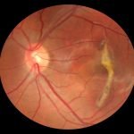


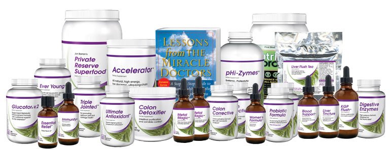

Hi, I was wondering what you
Hi, I was wondering what you meant by vitamin C should be taken as a complete complex?
The one antioxidant not
The one antioxidant not mentioned that is the most powerful is Astaxanthin. I was wondering what you think?
Respectfully,
Steve
As mentioned below,
As mentioned below, “Astaxanthin is oil based and is impossible to isolate from the oil. You either need to put it in its own capsule, or use it as a component in a large volume food. It requires too much volume to be part of a multi-antioxidant. Incidentally, Jon Barron has been using astaxanthin as part of his superfood formula since 2004. That’s long, long, long, long before it became the hot “new” antioxidant as touted in “many many” of today’s reports and research. It’s also a major component of krill oil, which Jon has also been recommending for many years.”
Hi Daniel,
To answer your
Hi Daniel,
To answer your question on vitamin C, check out this article: http://www.jonbarron.org/natural-health/barron-report-dietary-supplements-multivitamin
I've found articles on pubmed
I’ve found articles on pubmed indicating that restricting methionine and cysteine increase lifespans- at least in animals. So, I’m personally cautious about supplementing with those.
Hi,
My son got Cerebral Palsy
Hi,
My son got Cerebral Palsy and Epillepsy. What would be the best supplement or vitamins for him, especially to reduce his seizures? Thanks.
where is ASTAXANTHIN here
where is ASTAXANTHIN here ???
according to many many reports and research, Astaxanthin should be on top of all other antioxidants, including resveratrol.
Astaxanthin is oil based and
Astaxanthin is oil based and is impossible to isolate from the oil. You either need to put it in its own capsule, or use it as a component in a large volume food. It requires too much volume to be part of a multi-antioxidant. Incidentally, Jon Barron has been using astaxanthin as part of his superfood formula since 2004. That’s long, long, long, long before it became the hot “new” antioxidant as touted in “many many” of today’s reports and research. It’s also a major component of krill oil, which Jon has also been recommending for many years.
A “major component” of krill
A “major component” of krill oil ? Its in the MCG, and just around 100. That is peanuts. I have 12mg astaxanthin pills and trust me they do the job. Even 4mg is enough. Don’t spit out garbage. Astaxanthin is essential, never turning pro-oxidant…making it HUGELY BENEFICIAL in multi-oxidant roles. In my eyes, multi oxidant is an anomaly because they just lend each other electrons right ? I take Curcumin and Astaxanthin daily…its working great.
Dried fruit, including prunes
Dried fruit, including prunes, contain a carcinogenic chemical called acrylamide
Read more: http://www.livestrong.com/article/310301-prunes-diet/#ixzz2C7HexxMS
Please repond since I have replaced my “snacks” with prunes!
Does cooking veggies decrease
Does cooking veggies decrease their antioxidants? If so, is it substantial enough to be concerned about or change one’s eating habits?
Pardon me. I meant ultimate
Pardon me. I meant ultimate -antioxidant.
Isn’t it true that SOD taken
Isn’t it true that SOD taken orally does not get absorbed?
Yes and no. In the early days
Yes and no. In the early days, attempts to supplement with oral preparations of pure SOD enzyme proved disappointing, since the SOD protein molecule is easily deactivated by harsh acids and enzymes contained in the digestive tract. More modern versions of SOD supplements have conquered these challenges by creating bioavailable forms of SOD that are not neutralized by acids and enzymes.
what are the health benefits
what are the health benefits of CITRICIDAL liquid grapefruit seed extract? Does it work as an active antioxidant? I would appreciate your views.
Best regards
Elizabeth
Grapefruit seed extract is
Grapefruit seed extract is not an antioxidant; It is a broad spectrum anti-pathogen. At one time Jon used it in his anti-pathogen formula; however, he no longer does so. The primary reason is that it has become increasingly difficult to find “certified” food grade extract—even using the same sources that Citricidal does. In truth, the difference between industrial grade and food grade is not much – but it’s enough so that Jon no longer felt comfortable including it in the formula. It can certainly still be used externally for cleaning and disinfecting.
What I’m reading is that too
What I’m reading is that too much of a “good thing” may create other issues. I was taking a number of antioxidants and supplements. After some research, I discovered that quite a bit of my supplement regime fell into the category of DHT blockers, such as pygeum, nettles, green tea, peppermint, saw palmetto, zinc, grape seed extract, pumpkin seed, l-lysine, zinc, biotin, and lycopene, I have pretty good hair for a 65 year old male, but I began to notice a decreased sex drive,erectile dysfunction, and memory problems. Is there some way to supplement my diet with antioxidants without causing a decline in testosterone?
Yes, indeed. That’s one of
Yes, indeed. That’s one of Jon’s key messages. Too much a good thing can be bad. That said, be careful jumbling everything together. Your list is a mix of antioxidants, herbs, minerals, hormone regulators, and vitamins. Each one is different and has to be considered in its own right. That said, you might find the following link useful. http://www.jonbarron.org/article/30000-mile-tune
Dr. Baron,
Dr. Baron,
Have you heard of Protandim and what do you think about this “antioxidant” being a Nrf2 synergizer?
As a rule, Jon does not
As a rule, Jon does not comment on other companies products—especially network marketing products. (He receives, on average, about 600 such requests a month.) The quick comment on Protandim, though, is that it is very, very expensive for what it contains. You could buy the individual ingredients and take them for a lot less. Jon knows what the ingredients cost because he uses every one of them in several of his formulas. As to the proclaimed clinical studies supporting Protandim, there appear to be two such studies—a 2006 rodent study and a 2008 test tube study. That’s certainly interesting, but not particularly compelling. None of this is to say that it doesn’t work. If the raw materials used are high quality, they all can potentially provide benefits. It’s just that the final formula appears to be excessively expensive for what you get.
Thank you for yet another
Thank you for yet another comprehensive and competent article on antioxidants. I recently added Dr. P. Flanagan’s Hydrogen Boost to my antioxidant mix which contains most of the antioxidants you recommended in your article. As you didn’t mention negatively charged hydrogen ions bound to silica as powerful antioxidants I’m wondering what you, as an antioxidant specialist, could tell me about their proclaimed antioxidant effects. Is this just hype or is there some value in the argument about negatively charged hydrogen ions acting as antioxidants. Am I correct in assuming that they need to be synthetically produced and do not exist naturally that way? What would be their effect in the body? I would greatly appreciate, if you coud shed some light on this matter for me.
Negatively charged hydrogen
Negatively charged hydrogen ions bound to silica, as used by Flanagan are certainly a powerful antioxidant. But they still have the same issue all antioxidants have: they don’t work in all areas of the body, and they don’t work on all free radicals. You still need to think full-specturm.
What is your recommendation
What is your recommendation for the best antioxidant complex supplement?
Most of Jon Barron’s
Most of Jon Barron’s recommendations can be found on the “Products” link at the top of the page. For antioxidants, he links here: http://www.baselinenutritionals.com/products/ultimate-antioxidant.php
I take Jon Barron’s superfood
I take Jon Barron’s superfood formula, which the label says contains over 200 antioxidants. Is there any reason to take an antioxidant complex supplement too, or would that be redundant?
Remember, each antioxidant is
Remember, each antioxidant is different. Each one works on different free radicals and in different areas of the body. Depending on the antioxidant formula you take, it should make use of this fact by specifically targeting key free radicals and working in all key areas of the body. In other words, if the antioxidant formula you are taking is well designed and uses quality ingredients, it will not be redundant.
This is an amazing product.
This is an amazing product. Thank you for being here for us! I am wondering if you plan to use nicotinamide riboside in your formula…I have been reading that it’s great for anti aging and has been great as an antioxidant. I’m hesitant to take it though and would feel better if you were going to add it to any of your products.
Please let us know!
Thank you,
Nicotinamide riboside (NR) is
Nicotinamide riboside (NR) is a form of vitamin B3. And yes, a study with mice has proven interesting. As of yet, there is nothing definitive that shows it helps slow aging in humans—at least not in doses that are commercially available. In the mouse study, they supplemented the mice with a daily amount of 400 milligrams of NR per each kilogram of body mass. Translated to a human that weighs about 150 pounds, you’re looking at 28000 milligrams of NR per day, or 28 grams a day. Standard dosing for retail nicotinamide riboside is typically 250mg a day. Some people say that you shouldn’t use a simple weight conversion between mice and humans and that the actual dose need for humans is about 2200 mg. But that’s still 10 times the average daily supplement dose. Also, as Jon has pointed out in previous newsletters, only about 4-20% of mouse studies statistically translate to humans. The bottom line is that Jon finds the studies interesting, but doesn’t believe, at least at this point in time, that the science supports the use of NR as an antiaging ingredient.
Hello,
Hello,
I read that in some cases, anti-oxidants exacerbate cancer (if one already has it), since it has something to do with oxalates, and that free radicals can destroy cancer cells too.
This is confusing. What to do?
The answer is yes and no.
The answer is yes and no. Here is a brief excerpt from one of Jon’s articles on the topic. Just do a search on our site and you can read more: “
The Important Antioxidant Exception
When it comes to free radicals and antioxidants, cancer doesn’t follow the standard rule book. We already know a bit about antioxidants and free radicals, but to understand the problem here, we need to learn a bit more about the p53 gene.
Bottom line: cancer cells are capable of turning your body’s free radical/antioxidant systems upside down. They can co-opt antioxidants to protect themselves from free radicals that might kill them. Or they can allow antioxidants to throttle back your p53 gene, which then allows the cancer to multiply like crazy. This leaves us with a conundrum. If you stop using antioxidants before you get cancer, you might be increasing your odds of getting it in the first place. But if you’re using those same antioxidants in the early stages when you first get that cancer, you might be accelerating its growth unbeknownst to you. And if you stop using antioxidants with cancers that are susceptible to those antioxidants, you’re increasing your chances of succumbing to the cancer. What to do? What to do?
But here’s where the twist that I mentioned comes in–something not mentioned in any of the studies we’ve looked at today.
The Crucial Antioxidant Twist
Not all antioxidants are the same. They are as different as people at a UN meeting. Although, they’re all people, they look different; they act different, have different skills, and talk different languages. Likewise, different antioxidants work on different free radicals; they work in different parts of the body; and they work through different mechanisms. Some protect against aging. Some protect against cardiovascular damage. And some protect against cancer. Anyone making a blanket pronouncement on all antioxidant supplementation based on one study, on one kind of cancer, using one kind of antioxidant–and a synthetic or inorganic one at that–is practicing bad science and doing your health a disservice.
In summary, let’s take a quick look at how understanding this “twist” reveals how flawed the above studies’ recommendations concerning cancer and “ALL” antioxidants and “ALL” multivitamin combinations might be.
We could go on and on talking about studies that support the cancer protective effects of antioxidants such as curcumin, resveratrol, glutathione, ellagitannins, OPCs, etc.–not to mention the benefits of a number of vitamin combinations including C, D, and many of the B vitamins. But we’ve examined enough to demonstrate the problem making blanket conclusions based on limited studies of antioxidants and vitamins–especially when what’s being studied is synthetic or inorganic.”
Hello, after reading your
Hello, after reading your awesome list of antioxidants, I am interested in taking Alpha and Beta Carotene, L-Methionine, Green Tea Extract(I’ve taken before), Catalase, SOD and Astaxanthin(I’ve taken before, now again). Do you think taking all of these together is a good idea? I want to take them for their different properties that they exhibit. I also have Gingko Biloba 120 mg that I took on and off. I still have the bottle just haven’t been taking it as much as I used to. Thank you so much! This is the list I use whenever I want to decide to buy antioxidants.
Absolutely! That is why Jon
Absolutely! That is why Jon Barron even created a formula that essentially puts it all together for you, in the amounts you need to work together for maximum benefit. Most of Jon Barron’s recommendations can be found on the “Products” link at the top of the page. For antioxidants, he links here: https://Www.baselinenutritionals.com/Products/Ultimate-Antioxidant.php. Of course, you can create your own too, but be careful of sourcing each ingredient, the quality control of each company, and make sure the ingredients are in the right amounts to have any real health benefit.
Your article is very
Your article is very informative and it has helped me considerably in the search for suitable antioxidant sources to include in my diet. Thanks for writing it.
You say “Curcumin and green
You say “Curcumin and green tea strongly reinforce each other” in your article. Why is this, does the green tea enhance the absorption of the curcumin? Can you explain how this works?
According to a 1998 study
According to a 1998 study published in Carcinogenesis, varying concentrations of curcumin from Curcuma longa (turmeric) and epigallocatechin-3-gallate (EGCG) from Camellia sinensis (green tea) were incubated with four different human cell lines. One line was from normal tonsillar tissue, one from a precancerous lesion, and two from cancers. Unlike curcumin, EGCG had more difficulty impairing growth of the cell lines the more malignant they were. The combination of the two in a 1:1 ratio was significantly more effective at limiting cell growth than either alone. Flow cytometry studies showed that curcumin and EGCG had different mechanisms of action based on the fact that EGCG blocked cells at the G(1) phase of the cell cycle compared to G(2)/S for curcumin. In the normal cell line, the combination of curcumin and EGCG actually showed mild support of growth, indicating they were not toxic to healthy cells. The authors conclude that a combination of EGCG and curcumin may have significant, synergistic activity in preventing and reversing cancer. http://citeseerx.ist.psu.edu/viewdoc/download?doi=10.1.1.326.3618&rep=rep1&type=pdf
Great article, thank you for
Great article, thank you for your expertise. Wouldn’t it be best for all of us to have a yearly cancer check!!!??? Im a Celiac 23 yrs, been very ill since March with brain fog, depression, smells bothered me, couldnt eat, then insomnia. No help from several doctors who couldnt find what was wrong and charged me thousands! I discoverd I have malabsorption, and have healed the depression and much of the brain fog (still have some indecision etc.) – all with finding i had b-12 and d3 deficiency. I also have a suspicion that its cross reactive foods – that put me in this state….(age 49) because way back when I got Celiacs (26 but had it mildly before this for sev years) They did not know enough about healing the gut. So I researched and have been healing my gut, even going on SCD diet recently (and im seeing some improvement) I want to take some anti-oxidants in fear that with my body being so sick inside since March but prob well before that -(as i had depression 3 other times in the last 8 years and never knew it was all bcause of the gut health) that I wish to protect it from cancer. But how do I know if I already have cancer. I wish there was some test we could do annually !! I really need help for the insomnia, its going to take some time to start absorbing nutrients from food, as healing has to take place. But Im really wanting protection. If you have any advice, on the insomnia, (and I do take calcium/magnesium because I was def in calcium and i know how good mag is for you) but still no sleep. I end up taking unisom for a while and now its starting to not work. the back of my head hurts from lack of sleep (isnt that where your sleep center part of your brain is located)?
Unfortunately, there is no
Unfortunately, there is no single test for cancer. Each type of cancer requires its own test. You would have to run multiple yearly tests just for cancer to answer your question—and you would still not identify all the different types of cancer that you might get.