In our last newsletter, we explored the elite half of the immune system: cell-mediated immunity. These are the T-cells and Cytotoxic NK killer cells. To use a military analogy, these are the officers of the immune system — those educated at West Point. In this issue, we explore humoral immunity, the grunts of the immune system, the draftees who man the front lines and do the bulk of the fighting. These are primarily the B-cells, antibodies, phagocytes, granulocytes, and dendritic cells. In poetic terms, these are Tennyson’s “Six hundred.”1Alfred, Lord Tennyson. “The Charge of the Light Brigade.” eserver.org. Accessed 24 August 2011 This is where millions and millions of “cellular soldiers” are thrown into battle and sacrificed in defense of your body. And frequently, this is where the battles of life and death are fought — your life and death, that is.
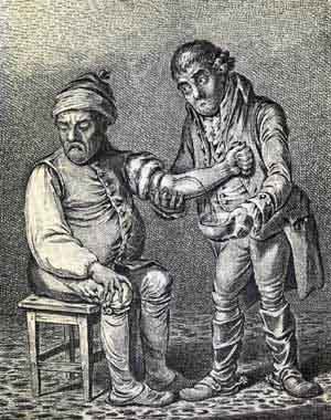
The word humoral is a throwback to the earliest days of medicine when doctors thought the body was regulated by four basic fluids called humors. Specifically, they thought that the body was filled with black bile, yellow bile, phlegm, and blood, and that illness occurred when these humors were out of balance. “Blood letting,” was a common treatment used to put the humors back in balance by reducing excess blood, thus bringing the body back into balance.
Nowadays, when doctors refer to “humoral immunity,” they’re just talking about that part of the immune response that takes place within body fluids (notably blood and lymph), not inside body cells, which, as we saw in the last newsletter, is where cell-mediated immunity works. In other words, humoral immunity works against extracellular pathogens in the blood and lymph — most notably bacteria and viruses not yet in cells, or that have been forced out of cells by cytotoxic NK cells.
Humoral (antibody-mediated) immunity
Humoral immunity targets invaders directly. To clarify, cell-mediated immunity defends against viruses that have taken up residence inside host cells and are doing their dirty deeds while ensconced there, whereas humoral immune cells take on invaders (bacteria, inactive viruses, fungi, molds, etc.) that are out in the open, traveling about in your blood and lymph. Humoral immune cells include:
- B-cells
- Antibodies
- Phagocytes
- Granulocytes
- Monocytes
- Dendritic cells
B-cells
When talking about humoral immunity, B-cells are at the head of the pack. The “B” in B-cells is now generally understood to refer to the “B” in bone marrow, where they are generated. B-cells travel directly from the bone marrow into your bloodstream. They do not pass go; they do not collect $200; and they do not go to the thymus like T-cells for specialized training. In fact, each B-cell comes out of the bone marrow programmed to make one specific antibody to defend against one specific invader. An antibody, by the way, is a soluble protein produced by B-cells that’s capable of binding to and destroying or neutralizing one particular foreign substance (antigen) in the body. Antibodies belong to a particular family of nine proteins called the immunoglobulins.
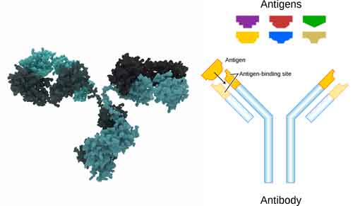
Antibodies in our immune system react with specific antigens to kill or neutralize them. They have a specific geometry that is crucial to their function. It is not quite like the lock and key mechanism of the T-cells, but it does include geometry specific antigen binding sites that grab onto and bind to specific antigens, as implied in the illustration above. So, one B-cell, for example, produces one particular antibody to defend against one particular strain of flu, whereas an entirely different B-cell produces the antibody for the strep bacteria, and so on. Note: there is a subset of B-cells that is a bit less specific and can work against multiple antigens. In fact, there are several subsets of B-cells, but four are primary.
- B2-cells are the specific antigen cells that we primarily think of when referring to B-cells, and will be the primary B-cells we discuss below.
- B1-cells have an affinity for multiple antigens. They are present in much fewer numbers than B2-cells and are found primarily in the peritoneal and pleural cavities.
- Plasma cells are the large cells produced by B-cells that have been exposed to a particular antigen. They are short lived — about one week — and die off quickly once no longer needed.
- Memory B-cells are formed from “activated B-cells” — B-cells that have been exposed to an antigen and finished attacking it. These function much like memory T-cells in that they are specific to the antigen encountered during the primary immune response. Memory B-cells live for a long time and, like memory T-cells, can respond quickly following a second exposure to the same antigen.
A critical difference between B-cells and T- cells is how each lymphocyte recognizes the antigen they’re geared to seek. As we discussed in Part 1 of our series, since T-cells have to identify invaders “hidden” inside body cells, they don’t get to look for the entire antigen. Instead, they have to recognize their targeted invader in a “processed” form, as a peptide fragment (a protein molecule) presented by the MHC molecule on the cell’s surface — the key to the T-cell’s lock, as it were. To learn this skill, they are trained in the thymus. B-cells, on the other hand, have an easier task. They get to look for their targeted antigen in its complete and unprocessed native form. They don’t have to look for bits and pieces of viral proteins, for example, they get to examine the complete target in all its glory. They get to track down free (soluble) antigens in the blood or lymph using their BCR (B-cell receptor) or membrane bound-immunoglobulin. To give you an analogy; it’s like playing “Name that Tune.” B-cells get to listen to the entire song before having to identify it; T-cells have to “name that tune” in just three notes.2Name that Tune. <http://www.youtube.com/watch?v=dWPvZI-ZnfQ> Much harder!
As mentioned previously, B-cells work primarily in the blood and lymph, defending against “foreign” invaders and toxic molecules. (In general, they are not as good at defending against the body’s own cells that have “gone bad” as T-cells, although in some cases, antibodies can indeed attack cancer cells.) By themselves, B-cells cannot produce a sufficient number of antibodies to overwhelm an invader. Instead, once a B-cell encounters the particular invader that it is built to defend against, it produces a vast number of plasma cells to manufacture the antibodies for them. Plasma cells are large cells that are essentially “factories” that produce millions of specific antibodies and release them into the bloodstream. Every single clone of the original plasma cell produced from a specific B-cell secretes a specific antibody for a single antigen. It is extremely specific. Each antibody targets one specific antigen and one only. Once the invader has been eliminated, the B-cells stop production of the plasma cells, and in a short period of time, most of the antibodies fade away.
Interestingly, even though they “work” in body fluids, B-cells do not generally circulate in the blood or lymph. Instead they camp out in the lymph nodes, spleen, and intestinal lymphatic tissue — waiting to be called into action. Like T-cells, they require costimulation to start working. The first stimulation comes from antigen receptors on their surfaces. They are activated by recognition of a specific antigen. The costimulation comes from interleukins (interleukin 2, 4, and 5) dispensed from T-helper cells. Once an invader has been recognized and the B-cells have been costimulated by a T-helper cell, they differentiate into plasma cells. As I mentioned previously, plasma cells function as antibody factories and secrete antigen-specific antibodies at a rate of millions of millions of molecules per cell per second. This is a major, major immune event/response.
Antibodies
Antibodies are Y-shaped protein molecules produced by B-cells that function as your body’s primary immune defense. Compared to the other components of the immune system, they are tiny. However, since they are proteins, they are also remarkably complex and composed of hundreds of amino acids folded over and over on each other, ultimately forming two chains. Their numbers dwarf by many magnitudes the numbers of T-cell defenders and macrophages. Each antibody molecule and its clones has a unique binding site that is set to combine with the complementary site of a foreign antigen. In the image below, on the left, you can see a representation of the complex folded amino acid structure of the antibody. On the right, there’s a representation of how the unique binding sites work.
Unlike T-cells, antibodies do not “actively” defend. They do not penetrate cell walls. They do not “consume” invaders, nor do they inject them with protein toxins. There is no “kiss of death” as seen with T-cells. Like Lilliputians taking on Gulliver, antibodies defeat invaders by sheer force of numbers.
Antibodies work by physically attaching themselves to the surface of an invading cell in massive numbers. The medical term for this is agglutination, which is defined exactly as it sounds (a gluing together). This has several possible effects on invaders such as bacteria. First, as more and more and more and more antibodies “agglutinate” to the bacteria, it becomes heavier and heavier until it literally precipitates out of the body fluid in which it is floating. At that point, it becomes easy prey for roving macrophages looking for detritus to eat up. The medical term for “coating” bacteria and other cells and rendering them subject to phagocytosis (being engulfed by phagocytes) is opsonization.
A secondary effect of massive agglutination is that the antibodies eventually build up to the point where they immobilize ciliated or flagellated bacteria. Once immobilized, they become easy targets for the macrophages. Ciliated bacteria are those that move about by virtue of all the little hairs on their surface, like the oars on a Roman galley. Flagellated bacteria, on the other hand, move about through the rotating motion of a tail-like structure that moves them forward like the propeller on a boat. When covered with antibodies, both processes come to a halt and the bacteria are immobilized.
Agglutination also allows antibodies to neutralize toxins from bacteria, such as tetanus, by literally encasing the toxins. Tetanus toxoid immunization injections, incidentally, artificially stimulate antibody production before exposure. (Immunization by vaccination is a controversial topic in the alternative health community, to say the least. We’ll talk more about immunization, the pros and cons, later on in our series on the immune system.)
And finally, antibodies serve as a form of preventative care in that they can stop viruses before they have a chance to enter cells, do their damage, and replicate exponentially.
Note: doctors often make use of antibodies’ specificity for particular antigens by cloning them in the laboratory to target specific antigens for either diagnosis, the delivery of chemicals, or even the ability to precisely delivery controlled doses of radiation. Antibodies cloned in the laboratory for these purposes are known as monoclonal antibodies.
Phagocytes
Most people are not aware of what phagocytes are or exactly what they do; nevertheless, when they think of the immune system, if they think of it at all, phagocytes are probably the cells they are thinking of. Phagocytes are the large white cells that eat and digest invading pathogens, primarily through protease enzyme activity. There are several kinds of phagocytes:
- Macrophages
- Neutrophils
- Monocytes
The word macrophage translates from the original Greek as “big eater.” And in fact macrophages are among the larger cells of the immune system and have a number of functions. Not only do they attack foreign invaders, they also play a key role as scavengers by “eating up” worn out cells and other waste in the body — referred to as necrotic tissue — especially in the lungs. Once macrophages have “digested” an invader, they then present the key identifying molecules, or antigens, to the T-cells to initiate the immune response.
From an alternative health point of view, macrophages play a key role in fasting. When you are not eating and creating new waste in the body, macrophages get a chance to “get ahead of the game” in terms of cleaning up debris. Fasting time thus becomes “spring cleaning” time for macrophages. Incidentally, macrophages look like amoebas, flowing from point to point, as they move about in pursuit of “enemies.” On another note, macrophages can play a significant role in the destruction of metastases in the early phases of tumor development, at least if your immune system is functioning properly. Unfortunately, at a certain point, tumors usually develop the ability to shut down macrophage attacks, thus ensuring their own survival. As always, it is easier for your immune system to prevent cancer from taking root than to eliminate it once it has. It of course can be done, but it takes much more effort and is more problematic.3Whitworth PW, Pak CC, Esgro J, Kleinerman ES, Fidler IJ. “Macrophages and cancer.” Cancer Metastasis Rev. 1990 Feb;8(4):319-51. <http://www.ncbi.nlm.nih.gov/pubmed/2182211>
Neutrophils, which are classed as both phagocytes and granulocytes, are actually the most abundant white cell in the body, comprising about 50% to 60% of the total circulating white blood cells, with each liter/quart of blood containing about five billion neutrophils. They have a very short life span, only living about six hours after leaving the bone marrow. However, it only takes them about 30 minutes to reach the site of an infection once they are released. Neutrophils rapidly attack any microorganisms coated with antibodies and kill them by eating them up, poisoning them with granule proteins, or capturing them in ultrafine nets created from their own DNA. Very cool! Once they have finished their work, they die and become the prime constituent of pus, along with captured bacteria, tissue debris, and blood serum.
At certain stages of cancer development, at least for some tumors, neutrophils can help fight cancer.4Fady C, Reisser D, Martin F. “Non-activated rat neutrophils kill syngeneic colon tumor cells by the release of a low molecular weight factor.” Immunobiology. 1990 Aug;181(1):1-12. <http://www.ncbi.nlm.nih.gov/pubmed/2272641>,5Blanks MJ, Stehle JR Jr, Du W, Adams JM, Willingham MC, Allen GO, Hu JJ, Lovato J, Molnar I, Cui Z. “Novel Innate Cancer Killing Activity in Humans.” Cancer Cell Int. 2011 Aug 3;11(1):26. [Epub ahead of print] <http://www.ncbi.nlm.nih.gov/pubmed/21813015> However, once tumors are established, certain types of tumors, such as breast cancer, don’t just shut down neutrophils, they can actually co-opt them to promote the growth and invasive nature of that tumor.6Marisa M. Queen, Randall E. Ryan, Ryan G. Holzer, Cynthia R. Keller-Peck, and Cheryl L. Jorcyk. “Breast Cancer Cells Stimulate Neutrophils to Produce Oncostatin M: Potential Implications for Tumor Progression.” Cancer Res October 1, 2005 65; 8896 <http://cancerres.aacrjournals.org/content/65/19/8896.full>,7Alyssa D. Gregory1 and A. McGarry Houghton. “Tumor-Associated Neutrophils: New Targets for Cancer Therapy.” Cancer Res April 1, 2011 71; 2411. <http://cancerres.aacrjournals.org/content/71/7/2411> Once again, it easier for the immune system to prevent cancer than to reverse it once it’s established. Once cancer is established, you need to be more selective in how you use the immune system to reverse it.
Monocytes are sort of the reserve, undifferentiated cells of the immune system. They don’t actually do much of anything themselves. Their importance lies in what they can become. They circulate in the blood for about three days after creation and then go hang out in different areas of the body, with about 50% being stored in the spleen, waiting to be called into action. As required by the nature of the infection or inflammation, they will flock to the area under attack and differentiate into macrophages and dendritic cells — thus providing a huge influx of warriors as needed.
Granulocytes
Granulocytes are named after the granular texture of their cytoplasm, which needless to say is granular. They include neutrophils (which function like phagocytes, but have the granular texture of granulocytes), eosinophils, basophils, and mast cells. Granulocytes destroy invaders by releasing granules filled with potent chemicals. Eosinophils play a key role in the killing of parasites through several specialized, highly toxic proteins present in the granules they release, including toxic basic protein and cationic protein. Basophils aren’t so much killers in their own right; they act more as facilitators. They release histamine and prostaglandins at the site of infection, which creates increased permeability of the tissue in the area, thus allowing for easier phagocyte migration to the site of infection so that the phagocytes can consume the invading microbes. In a normal, healthy immune system, granulocytes will aggressively kill certain types of cancer cells — at least until such time as the cancer figures out how to shut down the immune system. See for yourself.
Dendritic Cells
Dendritic cells have long threadlike tentacles that are used to wrap up antigens and expended lymphocytes and carry them to the lymph nodes for removal from the body. However, their primary function is to identify antigens, process them, and present them to the T-cells and B-cells to initiate the immune response.
Boosting your immune system
Now that we’ve slogged through a number of the details about the immune system, it’s time to collect the first piece of our reward — a discussion of how to actually boost immune function. And since we now have some shared background in the science behind immune function, we can do more than just list magic pills and herbs that you need to take on faith; we can actually discuss why and how they work — and work they do. Scientists have known for years that it is possible to improve the functioning of your immune system. The conventional medical approach has been to use expensive, proprietary drugs, including concentrated cytokines such as interleukin and interferon. Alternative healers, on the other hand, have adopted a more nuanced approach using natural substances to:
- Stimulate and strengthen the immune system
- Fight infection
- Strengthen tissue against assault by invading microorganisms
- Stimulate macrophage capability
- Increase T-cell production and protect helper T-cells
- Complement the action of interferon and interleukin-1
- Promote increased production of cytokines
- Assist the cell-mediated immune response
With that in mind, let’s take a look at some natural immune boosters. Not only are natural immune boosters safer than their pharmaceutical alternatives (having fewer side effects), they are, surprisingly, often more powerful– at least up to this point in time.
Echinacea
 There are several different ways that immune boosters can power up your immune system. One of the simplest is by presenting your immune system with what it perceives as a non-specific threat — a foreign antigen — that in actuality offers no real threat to the body. This causes your immune system to “power up” its defenses. However, since the immune booster presents no actual threat to the body, the immune system has nothing to use its new found readiness against. And thus it waits, charged up, primed for some/any threat to manifest so that it can jump on it with a vengeance. One thing to keep in mind about this kind of immune booster is that the immune system can be fooled by a false threat for only so long before it says to itself, “Ah, you’re just yankin’ my chain. I’m onto what’s happening here — time to stand down.” And thus the supplement stops working. When using immune boosters of this type, it’s best to use them for three weeks on and one week off. By taking that one week off, the immune system quickly forgets the false threat presented to the immune system. Thus, you can pull its leg again and again, while keeping your immune system on high alert indefinitely. You can do this because no memory cells are produced by the immune system since the immune system never actually gets to take the final step of “attacking” the immune booster, which is required for production of memory cells.
Note: if someone is highly sensitive to the antigens presented by this type of immune booster, their immune systems can actually “kick over” into an actual allergic response to the immune booster and produce symptoms such as sneezing and watery eyes, for example. For sensitive people, then, this type of immune booster is not useful. It should also be noted that this type of response can be plant part dependent. With Echinacea, for example, more people are sensitive to supplements made with Echinacea flowers as opposed to Echinacea seeds and roots. Fortunately, the strongest bioactives are in the Echinacea seeds and roots, not the flowers.
Echinacea (purple coneflower) was “discovered” in the late 1800’s by a traveling salesman named Joseph Meyer, who learned about it from the Plains Indians while traveling out West. He brewed it up as an alcohol tincture and sold it as a cure all — demonstrating its effectiveness by drinking his tonic and letting rattlesnakes bite him. Needless to say, he never got sick, from whence comes the phrase “snake oil.”
How does Echinacea work? In addition to tricking the immune system to ramp up, Echinacea helps in several other ways. First, it contains echinacoside, a natural antibiotic comparable to penicillin in effect, which can kill a broad range of viruses, bacteria, fungi, and protozoa.8Stoll, A., A. Renz & A. Brack. Antibacterial substances II. Isolation and constitution of echinacoside, a glycoside from the roots of echinacea augustifolia. Helvetic Chimica Acta, 33, 1877-1893, 1950. This makes it invaluable in wound healing and in the treatment of infectious diseases. Research has also reported Echinacea’s efficacy in treating colds, flu, bronchitis, and tuberculosis. Echinacea also contains echinacein, a biochemical that protects against germ attack by neutralizing the tissue-dissolving enzyme hyaluronidase, produced by many germs. Among the many pharmacological properties reported for Echinacea, the one demonstrated most convincingly is macrophage activation — by increasing production of interferon gamma.9Barrett B., Medicinal properties of Echinacea: A critical review, Phytomedecine, 2003, vol. 10, no1, pp. 66-86 [21 page(s). <http://www.ncbi.nlm.nih.gov/pubmed?term=12622467 > In addition, one study showed that Echinacea extracts can boost T-cell production by up to 30 percent more than immune boosting drugs.10Wagner H, Proksch A. An immunostimulating active constituent from Echinacea purpurea. Z Phytother 1981;2:166-171. Echinacea also increases production of the chemokines interleukin-8 and MCP-1, which enhance the migration of immune cells to the site of infection.
There are two primary varieties of Echinacea: purpurea and angustifolia. They are similar, but also have complementary properties. Formulas that use both are more likely to be effective. It’s also worth noting that potency runs from seed to root to leaf to almost none in the flower. And of course herb quality is paramount.
Over the last few years, there have been several studies that claimed to debunk Echinacea’s ability to boost the immune system and fight colds. Suffice it to say that the studies were either flawed in design (reviews of previously flawed studies), used the wrong parts of the Echinacea plant (flowers and leaves rather than roots and seeds), or used it at the wrong strength. A more recent study (2010), however, conducted using good quality Echinacea at a significant dose, found little benefit to using Echinacea in terms of reducing the length of a cold.11Bruce Barrett, MD, PhD; Roger Brown, PhD; Dave Rakel, MD; Marlon Mundt, PhD; et al. “Echinacea for Treating the Common Cold – A Randomized Trial.” Annals of Internal Medicine. December 21, 2010 vol. 153 no. 12 769-777. <http://www.annals.org/content/153/12/769.abstract> Not surprisingly, the press jumped all over it, proclaiming Echinacea was now proven to be little more than a placebo. However, two aspects of the study’s protocol negate the results.
- Dosing with Echinacea commenced at the onset of symptoms. This is too late to capitalize on Echinacea’s primary ability to ramp up the immune system. Once symptoms start, your immune system is going to be responding to the antigens presented by the cold virus so adding Echinacea will provide little added immune benefit at that point. (Remember, the key to Echinacea is ramping up the immune system “before” the invader arrives.) Any benefit will come from its germ killing properties, which although real, are secondary. And in that regard, the Echinacea did shorten the duration of colds — just not by that much. In truth, the major benefit of Echinacea is in its ability to prevent you from getting a cold in the first place — not shortening its duration — if you’ve been using it to build up your immune system in advance of being exposed to the virus.
- If you are going to wait until the last second, you have to intervene during the incubation phase at the latest, before symptoms fully manifest. And, at least with Echinacea, you have to use a liquid extract for quicker absorption. Once you hit the incubation phase, it’s only a matter of hours before the virus kicks into full gear. Waiting for an Echinacea pill to dissolve and make its way through the digestive tract takes too long. (We’ll talk more about the incubation phase when we talk about pathogen destroyers in Part 3 of our series.)
Forget the negative studies. Echinacea still stands as a powerful immune booster.
Pau d’arco
Pau d’arco (Tabebuia avellanedae, impetiginosa, and heptaphylla) is a tree that comes from the rain forests of Brazil and other areas of South America. It is the inner bark of the tree that provides the medicinal function.
Like Echinacea, this amazing herb both stimulates the body’s defense system and actively attacks pathogenic organisms. It has been used for centuries to improve immune function, detoxify, and reduce pain throughout the body, especially in the joints. Research has shown that it contains a natural antibacterial agent, has a healing effect on the entire body, cleanses the blood, and kills viruses. Pau d’arco has been used as a treatment for AIDS, allergies, infections and inflammations, anemia, asthma, arthritis and rheumatism, arteriosclerosis, bronchitis, cancer, candidiasis, colitis, cystitis, diabetes, eczema, fistulas, gastritis, gonorrhea, hemorrhages, Hodgkin’s disease, liver disease, leukemia, lupus, multiple sclerosis, osteomyelitis, Parkinson’s disease, prostatitis, psoriasis, skin sores, snake bites, ulcers, varicose veins, warts, and wounds.
The primary active biochemicals in Pau d’arco are the naphthoquinones: lapachol and beta-lapachone. Researchers have shown that lapachol has antitumorous, antiedemic, anti-inflammatory, antiseptic, antiviral, bactericidal, and antifungal activity.12Tandon VK, Singh RV, Yadav DB. Synthesis and evaluation of novel 1,4-naphthoquinone derivatives as antiviral, antifungal and anticancer agents. Bioorg Med Chem Lett. 2004 Jun 7;14(11):2901-4. <http://www.ncbi.nlm.nih.gov/pubmed/15125956>
Suma
Natives of the Amazon jungle have used suma root (Pfaffia paniculata) for at least the last 300 years. It wasn’t until 1975, however, that Suma was tested at the University of São Paulo, Brazil. The studies concluded that although it was not a cure, suma nevertheless brought significant relief for cancer,13T Watanabe, M Watanabe, Y Watanabe, C Hotta. “Effects of oral administration of Pfaffia paniculata (Brazilian ginseng) on incidence of spontaneous leukemia in AKR/J mice.” Cancer Detect Prev. 2000;24(2):173-8. <http://www.ncbi.nlm.nih.gov/pubmed/10917139> diabetes, and gout sufferers, with no undesirable side effects. Since then, studies at the American College of the Healing Arts have indicated that consistent use of suma may help combat fatigue (including treatment of chronic fatigue and low-energy conditions), prevent colds and flu, speed healing, regulate blood sugar, and stimulate the sex drive.
The key working ingredients in suma are pfaffic acid (prevents the spread of various cell disorders), pfaffocides and other saponins (helps stop diseases already in progress), the plant hormones sitosterol and stigmasterol (prevent cholesterol absorption and improve blood circulation), allantoin (helps accelerate healing), and germanium. Suma has one of the highest concentrations of germanium sesquioxide (Ge-132, aka organic germanium) of any plant known. Discovered about thirty years ago, Ge-132 works much like Pau d’arco in that it stimulates production of interferon gamma, while at the same time activating cytotoxic natural killer cells and macrophages. The net result is that it can invigorate the body, restore sexual function, protect against miscarriages, heal burns, reduce pain, treat circulatory disorders, and shrink cancers, in addition to being a powerful immunostimulant.14Stephen A. Levine, Ph.D. “Organic Germanium A Novel Dramatic Immunostimulant.” Orthomolecular.org, (Accessed 27 Aug 2011) <http://orthomolecular.org/library/jom/1987/pdf/1987-v02n02-p083.pdf>
Medicinal Mushrooms
Many of the compounds found in reishi, maitake, and cordyceps mushrooms are classified as host defense potentiators: it is believed that combinations of these compounds target and strengthen the human immune system, as well as aid in neuron transmission, metabolism, hormonal balance, and the transport of nutrients and oxygen. Through a host-mediated (T-cell) immune mechanism, they help the body regulate the development of lymphoid stem cells and other important defense responses.
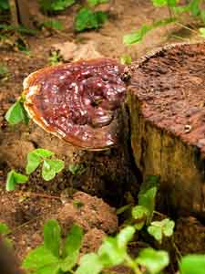 Reishi (Ganoderma lucidum or lingzhi) — The anti-cancer15Xu Z, Chen X, Zhong Z, Chen L, Wang Y. “Ganoderma lucidum polysaccharides: immunomodulation and potential anti-tumor activities.” Am J Chin Med. 2011;39(1):15-27. <http://www.ncbi.nlm.nih.gov/pubmed/21213395> and immune-enhancing effects16Watanabe K, Shuto T, Sato M, Onuki K, Mizunoe S, Suzuki S, Sato T, Koga T, Suico MA, Kai H, Ikeda T. “Lucidenic acids-rich extract from antlered form of Ganoderma lucidum enhances TNFa induction in THP-1 monocytic cells possibly via its modulation of MAP kinases p38 and JNK.” Biochem Biophys Res Commun. 2011 Apr 29;408(1):18-24. Epub 2011 Mar 29. <http://www.ncbi.nlm.nih.gov/pubmed/21453678> of the reishi mushroom are thought to be largely due to its mucopolysaccharides, which the body incorporates into cellular membranes, making them resistant to viruses and pathogenic bacteria and the triterpenes, which induce tumor necrosis factor production. The polysaccharides appear to activate macrophages that “consume” viruses, bacteria, and other large particulate matter.
Maitake (Grifola frondosa, also known as Sheep’s Head and Hen of the Woods) — Maitake mushrooms have a very high concentration of a unique polysaccharide compound called beta-1,6-glucan, which researchers consider to be one of the most powerful immune stimulants and adaptogens known. One study showed that maitake produced a 64 percent inhibition of breast cancer and tumor activity and a 75 percent inhibition of skin cancer and tumor activity.17Hiroaki Nanba, Ph.D. “Maitake D-fraction: Healing and Preventive Potential for Cancer.” The Journal of Orthomolecular Medicine Vol. 12, 1st Quarter 1997. <http://orthomolecular.org/library/jom/1997/articles/1997-v12n01-p043.shtml> <http://orthomolecular.org/library/jom/1997/articles/1997-v12n01-p043.shtml> Also, laboratory studies conducted at the U.S. National Cancer Institute (NCI) and the Japanese National Institute of Health showed that maitake extract kills the human immunodeficiency virus (HIV) and enhances the activity of helper T-cells.18Ishikawa, K. “Anti-HIV Activity in Cytopathic Effect of Proteoglucan Extracted from Maitake Mushroom,” National Institute of Health, Jan 23, 1991. In fact, the NCI researchers reported that the maitake extract was as powerful as AZT (a commonly prescribed AIDS drug) but without the toxic side effects.
Research has demonstrated that maitake stimulates the production of a variety of immune cells, including macrophages, NK cells, and T-cells, and it increases their effectiveness by increasing the production of interleukin-l, interleukin-2, and lymphokines. It also stimulates the bone marrow to produce stem cells and granulocytes by stimulating production of the cytokine granulocyte colony stimulating factor.19Ito K, Masuda Y, Yamasaki Y, Yokota Y, Nanba H. “Maitake beta-glucan enhances granulopoiesis and mobilization of granulocytes by increasing G-CSF production and modulating CXCR4/SDF-1 expression.” Int Immunopharmacol. 2009 Sep;9(10):1189-96. Epub 2009 Jun 30. <http://www.ncbi.nlm.nih.gov/pubmed/19573626> Further, maitake has been confirmed to have a multifaceted benefit for treating cancer and tumors: it protects healthy cells from becoming cancerous, helps prevent the spread of cancer (metastasis), and slows or stops the growth of tumors.20Kodama N, Mizuno S, Nanba H, Saito N. “Potential antitumor activity of a low-molecular-weight protein fraction from Grifola frondosa through enhancement of cytokine production.” J Med Food. 2010 Feb;13(1):20-30. <http://www.ncbi.nlm.nih.gov/pubmed/20136432> Maitake works in conjunction with chemotherapy by lessening the negative side effects (by as much as 90 percent).
Cordyceps has properties similar to those of ginseng and has been used to strengthen and rebuild the body after exhaustion or long-term illness. It is one of the most valued medicinal fungi in Chinese medicine. It has also been used traditionally for impotence, neurasthenia, and backache. Recent research with extracts of Cordyceps has yielded a protein-bound polysaccharide with activity against tumors, as well as being capable of upregulating macrophage activity,21Lee JS, Hong EK. “Immunostimulating activity of the polysaccharides isolated from Cordyceps militaris.” Int Immunopharmacol. 2011 Sep;11(9):1226-33. Epub 2011 Apr 14. <http://www.ncbi.nlm.nih.gov/pubmed/21497206> and inducing the apoptosis (cell death) of human leukemia cells.22Jeong JW, Jin CY, Park C, Hong SH, Kim GY, Jeong YK, Lee JD, Yoo YH, Choi YH. “Induction of apoptosis by cordycepin via reactive oxygen species generation in human leukemia cells.” Toxicol In Vitro. 2011 Jun;25(4):817-24. Epub 2011 Feb 15. <http://www.ncbi.nlm.nih.gov/pubmed/21310227> Cordyceps is widely employed to treat upper respiratory problems, impotence, and weakened immune systems, and also by athletes to increase endurance.
AHCC (Active Hexose Correlated Compound) — AHCC is a proprietary dietary supplement rich in polysaccharides and fiber derived from mushrooms. Studies have shown that it can be effective in stimulating the production of NK cells, killer T-cells, and cytokines (interferon, interleukin-12, and TNF-alpha). In Japan, it is used extensively in hospitals in combination with chemotherapy treatments to reduce the adverse side effects of those treatments.23AHCC Published Research. Accessed 28 Aug 2011. <http://www.ahccpublishedresearch.com/default.htm>
Astragalus membranaceus
Astragalus has been a foundational herb in Traditional Chinese Medicine for hundreds of years. It is one of the important “Qi tonifying” adaptogenic herbs from the Chinese materia medica. Current research on astragalus focuses on the immune stimulating capacity of its polysaccharides and saponins. It also appears to be useful in dealing with cancer,24Auyeung KK, Mok NL, Wong CM, Cho CH, Ko JK. “Astragalus saponins modulate mTOR and ERK signaling to promote apoptosis through the extrinsic pathway in HT-29 colon cancer cells.” Int J Mol Med. 2010 Sep;26(3):341-9. <http://www.ncbi.nlm.nih.gov/pubmed/20664949> and in increasing stamina. First and foremost, though, it is an immunostimulant25Liu QY, Yao YM. “The regulatory effect and mechanism of Astragalus polysaccharides on CD11c(high)CD45RB(low) dendritic cell.” Zhonghua Shao Shang Za Zhi. 2011 Apr;27(2):95-9. <http://www.ncbi.nlm.nih.gov/pubmed/21651844> used in the treatment of chronic viral infections, hepatitis, edema, common cold, and flu. Astragalus increases the interferon response to viral infection and works synergistically with interferon. It also increases phagocytic activity and antibody levels and improves the functioning of natural killer cells.
Aloe vera
The polysaccharide component of aloe vera, acemannan, possesses significant immune-enhancing and antiviral activity. Supplementing with acemannan has been proven to increase lymphocyte response to antigens by enhancing the release of interleukin-I. In addition, acemannan has been shown to increase macrophage levels and have a positive effect on T-cell activity and dendritic cell maturation.26Lee JK, Lee MK, Yun YP, Kim Y, Kim JS, Kim YS, Kim K, Han SS, Lee CK. “Acemannan purified from Aloe vera induces phenotypic and functional maturation of immature dendritic cells.” Int Immunopharmacol. 2001 Jul;1(7):1275-84. <http://www.ncbi.nlm.nih.gov/pubmed?term=11460308> Look for whole leaf aloe extract, which is two to three times more potent than gel/juice. Why? The greatest concentration of active ingredients is at the interface of the rind and the inner gel. If your extract doesn’t come from the whole leaf, you lose half to two-thirds of the active biochemicals.
 Alkylglycerols
Alkylglycerols (AKGs) are lipids naturally manufactured in the body and found in mother’s milk, the liver and spleen, and bone marrow. They play a major role in the production and stimulation of white blood cells. They also help to normalize bone marrow function. The immune-supportive effect of AKGs helps our bodies protect against bacterial, fungal, and viral infections through enhanced phagocytosis (eating up the bad guys) and antibody production.27Ngwenya BZ, Foster DM. ” Enhancement of antibody production by lysophosphatidylcholine and alkylglycerol.” Proc Soc Exp Biol Med. 1991 Jan;196(1):69-75. <http://www.ncbi.nlm.nih.gov/pubmed/1984244> The most potent source of AKGs in the world is shark liver oil.
Colostrum and Lactoferrin
Colostrum is the clear, yellowish, pre-milk fluid produced from the mother’s mammary glands during the first seventy-two hours after birth. It provides both immune and growth factors essential for the health and vitality of the newborn. Obviously, supplementation with human colostrum is not an option, but researchers have found that bovine colostrum (from cows) is virtually identical, except that the immune factors are actually several times more concentrated.
The immune factors in colostrum have been shown to help the body resist pathogens such as viruses,28”Clinical Tests: Preventing Colds with APS45-10 Colostrum.” Paper presented at Annual Meeting of Japanese Society of Clinical Nutrition 2007, The 13th Symposium of Adult Disease Countermeasure Society 2008, The 14th Symposium of Adult Disease Countermeasure Society 2009 and Annual Meeting of Japanese Dairy Science Association 2009. <http://www.apsbiogroup.com/colostrum/references/Preventing_Colds.pdf> bacteria, yeast, and fungi. In addition, colostrum contains a number of antibodies to specific pathogens, including E. coli, salmonella, rotavirus, Candida, streptococcus, staphylococcus, H. pylori, and cryptosporidia. Proline-rich-polypeptide, a component of colostrum, works as an immunomodulator, boosting a low immune system and balancing an overactive one. (We’ll talk more about immunomodulators in our next newsletter.) Another key component of colostrum is transfer factors, small molecules that transfer immunity information from one entity to another. In effect, they transfer immunity “memory,” thereby giving you instant resistance to a number of diseases.
Colostrum is a potent source of lactoferrin, a globular protein produced in the body. It is found anywhere that is especially vulnerable to attack, such as in the gut, eyes, ears, nose, throat, and urinary tract. Lactoferrin has been shown to inhibit virus replication (including AIDS and herpes viruses), limit tumor growth and metastasis, directly kill both bacteria and yeast (including Candida), and activate neutrophils. Supplementation with lactoferrin can significantly boost the immune system and help the body recover from any existing infection. Maintaining healthy levels of intestinal flora through the use of probiotic supplements allows the body to produce its own lactoferrin.
Look for colostrum obtained from organic, grass-fed dairy cows and standardized to 40% Immunoglobulins.
Glutathione
Glutathione is a tripeptide molecule found in human cells. In addition to being a powerful antioxidant, glutathione works to support the active functioning of the immune system and is a key component of all lymphocytes. In fact, all lymphocytes require sufficient levels of intracellular glutathione to function properly. It also plays a major protective role against the damaging effects of the whole range of pathogens and carcinogens. For many people, glutathione supplements are upsetting to the stomach. Alternatives include the glutathione precursors L-cysteine and L-glutamate and specially formulated whey products.
Mangosteen
Mangosteen (Garcinia mangostana) is a tropical evergreen tree whose contains a unique group of antioxidants called xanthones. Xanthones, particularly beta and gamma mangostin, work to maintain the immune system, support cardiovascular health, optimize joint flexibility, are naturally antibiotic, antiviral, and anti-inflammatory, and are some of the most powerful antioxidants found in nature. In addition, recent studies have confirmed that gamma mangostin is a potent COX inhibitor, an important factor in reducing inflammation, pain, and fever. Other studies have shown that alpha-mangostin can enhance the body’s innate responses to viral infection.29Shaneyfelt ME, Burke AD, Graff JW, Jutila MA, Hardy ME. “Natural products that reduce rotavirus infectivity identified by a cell-based moderate-throughput screening assay.” Virol J. 2006 Sep 1;3:68. <http://www.ncbi.nlm.nih.gov/pubmed/16948846> And as has been true with most of the other immune boosters we’ve looked at so far, mangosteen has also shown the ability to work as an anticancer agent. Specifically, the antimetastatic activity of alpha-mangostin has been demonstrated in clinical studies on breast cancer.30Shibata MA, Iinuma M, Morimoto J, Kurose H, Akamatsu K, Okuno Y, Akao Y, Otsuki Y. “a-Mangostin extracted from the pericarp of the mangosteen (Garcinia mangostana Linn) reduces tumor growth and lymph node metastasis in an immunocompetent xenograft model of metastatic mammary cancer carrying a p53 mutation.” BMC Med. 2011 Jun 3;9:69. <http://www.ncbi.nlm.nih.gov/pubmed/21639868>
Beta-glucan
Beta-glucan is a natural complex carbohydrate (polysaccharide) found in cereal grains such as oats and barley. However, it is found in its greatest concentration primarily in the cell walls of yeast and in medicinal mushrooms. Beta Glucan as a supplement, however, particularly Beta- 1,3/1,6 Glucan, extracted from yeast cell wall, is a potent and proven immune response potentiator and modulator — stimulating anti-tumor and antimicrobial activity by binding to receptors on macrophages and other white blood cells and activating them.31Beta Glucan Reasearch. <http://www.betaglucan.org/>
Conclusion
In this part of our series on the immune system we covered the humoral immune system (B-cells and antibodies), and explored some of the ways you can enhance your immune system. In addition, we learned that natural immune enhancement can follow several different routes, from tricking the immune system into thinking it’s under attack to increasing cytokine production to stimulating the production of immune cells to providing specific building blocks (such as glutathione) that your immune system needs to function.
One other important thing that we learned about is the connection between the immune system and cancer and how most immune boosters also function as anti-cancer agents, each helping your body fight cancer and metastasis in their own unique way. Also, by combining what we’ve learned over the first two parts of this series, we’ve gained an understanding of how the immune system actually does this. In simple terms, as genes in a human cell are damaged or mutate over time, eventually turning the cell cancerous, they signal that change in their internal structure by expressing different protein molecules on the cell’s surface. At a certain point, these changing surface proteins tell the immune system that the cancer cell is no longer “self”, but “non-self” and must be attacked. Obviously, this is not a flawless process, or no one would ever come down with cancer. But it is a process that can be encouraged, supported, and enhanced — both through natural and medical means — and in fact, a potentially revolutionary breakthrough in the treatment of cancer that was just announed does exactly that.32David L. Porter, M.D., Bruce L. Levine, Ph.D., Michael Kalos, Ph.D., Adam Bagg, M.D., and Carl H. June, M.D. “Chimeric Antigen Receptor–Modified T Cells in Chronic Lymphoid Leukemia.” N Engl J Med 2011; 365:725-733 August 25, 2011. <http://www.nejm.org/doi/full/10.1056/NEJMoa1103849> In a future newsletter series, when I revisit cancer, we’ll cover this subject in more detail.
In the next part of our series, we’ll explore:
- The complementary immune system
- Circulating immune complexes
- Natural immunomodulators
- Potential problems with the immune system
- Pathogen destroyers and the incubation phase
- How we build immunity
Click below if you’ve missed any part of this Immunity series:
Anatomy and Physiology of the Immune System, Part 1
Anatomy and Physiology of the Immune System, Part 2
Anatomy and Physiology of the Immune System, Part 3
Anatomy and Physiology of the Immune System, Part 4
References
| ↑1 | Alfred, Lord Tennyson. “The Charge of the Light Brigade.” eserver.org. Accessed 24 August 2011 |
|---|---|
| ↑2 | Name that Tune. <http://www.youtube.com/watch?v=dWPvZI-ZnfQ> |
| ↑3 | Whitworth PW, Pak CC, Esgro J, Kleinerman ES, Fidler IJ. “Macrophages and cancer.” Cancer Metastasis Rev. 1990 Feb;8(4):319-51. <http://www.ncbi.nlm.nih.gov/pubmed/2182211> |
| ↑4 | Fady C, Reisser D, Martin F. “Non-activated rat neutrophils kill syngeneic colon tumor cells by the release of a low molecular weight factor.” Immunobiology. 1990 Aug;181(1):1-12. <http://www.ncbi.nlm.nih.gov/pubmed/2272641> |
| ↑5 | Blanks MJ, Stehle JR Jr, Du W, Adams JM, Willingham MC, Allen GO, Hu JJ, Lovato J, Molnar I, Cui Z. “Novel Innate Cancer Killing Activity in Humans.” Cancer Cell Int. 2011 Aug 3;11(1):26. [Epub ahead of print] <http://www.ncbi.nlm.nih.gov/pubmed/21813015> |
| ↑6 | Marisa M. Queen, Randall E. Ryan, Ryan G. Holzer, Cynthia R. Keller-Peck, and Cheryl L. Jorcyk. “Breast Cancer Cells Stimulate Neutrophils to Produce Oncostatin M: Potential Implications for Tumor Progression.” Cancer Res October 1, 2005 65; 8896 <http://cancerres.aacrjournals.org/content/65/19/8896.full> |
| ↑7 | Alyssa D. Gregory1 and A. McGarry Houghton. “Tumor-Associated Neutrophils: New Targets for Cancer Therapy.” Cancer Res April 1, 2011 71; 2411. <http://cancerres.aacrjournals.org/content/71/7/2411> |
| ↑8 | Stoll, A., A. Renz & A. Brack. Antibacterial substances II. Isolation and constitution of echinacoside, a glycoside from the roots of echinacea augustifolia. Helvetic Chimica Acta, 33, 1877-1893, 1950. |
| ↑9 | Barrett B., Medicinal properties of Echinacea: A critical review, Phytomedecine, 2003, vol. 10, no1, pp. 66-86 [21 page(s). <http://www.ncbi.nlm.nih.gov/pubmed?term=12622467 > |
| ↑10 | Wagner H, Proksch A. An immunostimulating active constituent from Echinacea purpurea. Z Phytother 1981;2:166-171. |
| ↑11 | Bruce Barrett, MD, PhD; Roger Brown, PhD; Dave Rakel, MD; Marlon Mundt, PhD; et al. “Echinacea for Treating the Common Cold – A Randomized Trial.” Annals of Internal Medicine. December 21, 2010 vol. 153 no. 12 769-777. <http://www.annals.org/content/153/12/769.abstract> |
| ↑12 | Tandon VK, Singh RV, Yadav DB. Synthesis and evaluation of novel 1,4-naphthoquinone derivatives as antiviral, antifungal and anticancer agents. Bioorg Med Chem Lett. 2004 Jun 7;14(11):2901-4. <http://www.ncbi.nlm.nih.gov/pubmed/15125956> |
| ↑13 | T Watanabe, M Watanabe, Y Watanabe, C Hotta. “Effects of oral administration of Pfaffia paniculata (Brazilian ginseng) on incidence of spontaneous leukemia in AKR/J mice.” Cancer Detect Prev. 2000;24(2):173-8. <http://www.ncbi.nlm.nih.gov/pubmed/10917139> |
| ↑14 | Stephen A. Levine, Ph.D. “Organic Germanium A Novel Dramatic Immunostimulant.” Orthomolecular.org, (Accessed 27 Aug 2011) <http://orthomolecular.org/library/jom/1987/pdf/1987-v02n02-p083.pdf> |
| ↑15 | Xu Z, Chen X, Zhong Z, Chen L, Wang Y. “Ganoderma lucidum polysaccharides: immunomodulation and potential anti-tumor activities.” Am J Chin Med. 2011;39(1):15-27. <http://www.ncbi.nlm.nih.gov/pubmed/21213395> |
| ↑16 | Watanabe K, Shuto T, Sato M, Onuki K, Mizunoe S, Suzuki S, Sato T, Koga T, Suico MA, Kai H, Ikeda T. “Lucidenic acids-rich extract from antlered form of Ganoderma lucidum enhances TNFa induction in THP-1 monocytic cells possibly via its modulation of MAP kinases p38 and JNK.” Biochem Biophys Res Commun. 2011 Apr 29;408(1):18-24. Epub 2011 Mar 29. <http://www.ncbi.nlm.nih.gov/pubmed/21453678> |
| ↑17 | Hiroaki Nanba, Ph.D. “Maitake D-fraction: Healing and Preventive Potential for Cancer.” The Journal of Orthomolecular Medicine Vol. 12, 1st Quarter 1997. <http://orthomolecular.org/library/jom/1997/articles/1997-v12n01-p043.shtml> <http://orthomolecular.org/library/jom/1997/articles/1997-v12n01-p043.shtml> |
| ↑18 | Ishikawa, K. “Anti-HIV Activity in Cytopathic Effect of Proteoglucan Extracted from Maitake Mushroom,” National Institute of Health, Jan 23, 1991. |
| ↑19 | Ito K, Masuda Y, Yamasaki Y, Yokota Y, Nanba H. “Maitake beta-glucan enhances granulopoiesis and mobilization of granulocytes by increasing G-CSF production and modulating CXCR4/SDF-1 expression.” Int Immunopharmacol. 2009 Sep;9(10):1189-96. Epub 2009 Jun 30. <http://www.ncbi.nlm.nih.gov/pubmed/19573626> |
| ↑20 | Kodama N, Mizuno S, Nanba H, Saito N. “Potential antitumor activity of a low-molecular-weight protein fraction from Grifola frondosa through enhancement of cytokine production.” J Med Food. 2010 Feb;13(1):20-30. <http://www.ncbi.nlm.nih.gov/pubmed/20136432> |
| ↑21 | Lee JS, Hong EK. “Immunostimulating activity of the polysaccharides isolated from Cordyceps militaris.” Int Immunopharmacol. 2011 Sep;11(9):1226-33. Epub 2011 Apr 14. <http://www.ncbi.nlm.nih.gov/pubmed/21497206> |
| ↑22 | Jeong JW, Jin CY, Park C, Hong SH, Kim GY, Jeong YK, Lee JD, Yoo YH, Choi YH. “Induction of apoptosis by cordycepin via reactive oxygen species generation in human leukemia cells.” Toxicol In Vitro. 2011 Jun;25(4):817-24. Epub 2011 Feb 15. <http://www.ncbi.nlm.nih.gov/pubmed/21310227> |
| ↑23 | AHCC Published Research. Accessed 28 Aug 2011. <http://www.ahccpublishedresearch.com/default.htm> |
| ↑24 | Auyeung KK, Mok NL, Wong CM, Cho CH, Ko JK. “Astragalus saponins modulate mTOR and ERK signaling to promote apoptosis through the extrinsic pathway in HT-29 colon cancer cells.” Int J Mol Med. 2010 Sep;26(3):341-9. <http://www.ncbi.nlm.nih.gov/pubmed/20664949> |
| ↑25 | Liu QY, Yao YM. “The regulatory effect and mechanism of Astragalus polysaccharides on CD11c(high)CD45RB(low) dendritic cell.” Zhonghua Shao Shang Za Zhi. 2011 Apr;27(2):95-9. <http://www.ncbi.nlm.nih.gov/pubmed/21651844> |
| ↑26 | Lee JK, Lee MK, Yun YP, Kim Y, Kim JS, Kim YS, Kim K, Han SS, Lee CK. “Acemannan purified from Aloe vera induces phenotypic and functional maturation of immature dendritic cells.” Int Immunopharmacol. 2001 Jul;1(7):1275-84. <http://www.ncbi.nlm.nih.gov/pubmed?term=11460308> |
| ↑27 | Ngwenya BZ, Foster DM. ” Enhancement of antibody production by lysophosphatidylcholine and alkylglycerol.” Proc Soc Exp Biol Med. 1991 Jan;196(1):69-75. <http://www.ncbi.nlm.nih.gov/pubmed/1984244> |
| ↑28 | ”Clinical Tests: Preventing Colds with APS45-10 Colostrum.” Paper presented at Annual Meeting of Japanese Society of Clinical Nutrition 2007, The 13th Symposium of Adult Disease Countermeasure Society 2008, The 14th Symposium of Adult Disease Countermeasure Society 2009 and Annual Meeting of Japanese Dairy Science Association 2009. <http://www.apsbiogroup.com/colostrum/references/Preventing_Colds.pdf> |
| ↑29 | Shaneyfelt ME, Burke AD, Graff JW, Jutila MA, Hardy ME. “Natural products that reduce rotavirus infectivity identified by a cell-based moderate-throughput screening assay.” Virol J. 2006 Sep 1;3:68. <http://www.ncbi.nlm.nih.gov/pubmed/16948846> |
| ↑30 | Shibata MA, Iinuma M, Morimoto J, Kurose H, Akamatsu K, Okuno Y, Akao Y, Otsuki Y. “a-Mangostin extracted from the pericarp of the mangosteen (Garcinia mangostana Linn) reduces tumor growth and lymph node metastasis in an immunocompetent xenograft model of metastatic mammary cancer carrying a p53 mutation.” BMC Med. 2011 Jun 3;9:69. <http://www.ncbi.nlm.nih.gov/pubmed/21639868> |
| ↑31 | Beta Glucan Reasearch. <http://www.betaglucan.org/> |
| ↑32 | David L. Porter, M.D., Bruce L. Levine, Ph.D., Michael Kalos, Ph.D., Adam Bagg, M.D., and Carl H. June, M.D. “Chimeric Antigen Receptor–Modified T Cells in Chronic Lymphoid Leukemia.” N Engl J Med 2011; 365:725-733 August 25, 2011. <http://www.nejm.org/doi/full/10.1056/NEJMoa1103849> |


How can you balance out an
How can you balance out an over aggressive Immune system? Vitiligo is an autoimmune condition where the T cells begin killing off the pigment producing cells of the body. Is there any research done to verify this theory and , if so, are there any treatment available? Thanks. Stan Dass.
I have been a veterinary
I have been a veterinary medical doctor and researcher for almost 30 years. The vaccines are the largest contributor to dys regulated immune systems and unhealthy Th2 bias (humoral) immune system expression. My recent presentation on Vaccinosis showed vitiligo as just one of the many ways the vaccine induced disease can express. Only vaccinated populations have these high rates of auto immune diseases. The Hayward Study showed definitively that only vaccinated individuals were developing auto antibodies that then lead to autoimmune disease. Antibiotics and other drugs also increase autoimmune disease. Anyway, we are all right now trying to see if the vaccine damage can be reversed, one way that we have cured vitiligo is with homeopathic remedy thuja which is classically the largest remedy used and recognized for vaccinosis. Eating a plant based diet also is important as the processed foods, carbs, starches, sugars are very inflammatory. Raw diets that are full of vitmins, minerals and exzymes, cofactors etc are also necessary to decrease the overall inflammation and get what you can reversed with optimal nutrition. The most important thing in the world is for everyone to realize that vaccines are exactly the great immune impactor and not in a good way. The damage is cumulative, In animals that are so so highly over vaccinated their helath is ruined by 5-7 years if they are getting yearly vaccines. Our leading veterinary researcher, Dr. Ron Schultz has been on public record since 1978 reporting that only one lethal virus vaccine is necessary to convey a lifetime of immunity for a mature mammal. No vaccines to bacteria or to parasites are worth taking.
The vaccines impact our immune system and actually are responsible for the reaction and many times the reaction is the pathology of the disease. Our gene profile expression of disease is the result of vaccinations!Knowing now what I do about vaccines and our immune system and the whole body impact, I would never again.
Can you recommend any
Can you recommend any articles to read for those who are diagnosed with chronic lymphocytic leukemia?
Thanks,
R McFee
You can follow the link in
You can follow the link in footnote 32 to the original published study in the New England Journal of Medicine, or for a layman’s discussion of the study, check out:
http://www.medpagetoday.com/HematologyOncology/Leukemia/27997?pfc=101&spc=224.
I would start by going to our
I would start by going to our health topic on cancer: http://www.jonbarron.org/alternative-cancer/natural-health-remedies-immune-system. You can also do a search on leukemia at the top of this website and read all the articles that come up in the site search.
I can see the value in
I can see the value in Maitake and Reishi mushrooms, but how about the supermarket “white button” variety? Are they as healthful or just tasty?
Hi Jon. While the above
Hi Jon. While the above article is very interesting to read, it reflects "old type" thinking of the medical establishment. (pathogenic) viruses are a figment of the conventional medical establishment and have never, to this day been proven to exist. What is commonly referred to as viruses, are bits of protein, made by the body cells for the body to use as "building materials" to repair damage caused either through trauma (injury) or by a specific biological SPECIAL program of Nature. The so called viruses found on the cervix are a case in point. Cervical 'cancer' is such a PROGRAM of Nature, which has two phases to it. Phase one is an ulceration caused by the perception of the Psyche, that fertilization did not, or could not take place, constituting a biological conflict. Upon resolution of this conflict, phase two is initiated and the ulceration is being restored to normalcy, using said 'virus material'.
Yes, I am familiar with the
Yes, I am familiar with the cell detritus theory, but it is hardly new. It's roots go back to the 1920's with the work of Dr. Herbert Shelton and his disciple T.C. Fry. And as theories go, there is significantly more evidence that exists in support of the pathogenic virus theory than in support of the virus as cell detrius theory. If we are both around in another 50 years, we can compare notes and see which theory was finally validated. Incidentally, the first virus (rightly or wrongly) was identified in the 1880's — so only 40 years separates the two theories. In other words, both theories represent "old type" thinking. Sorry!
In the meantime, I can pretty much tell you that based on my observations of tens of thousands of people who have used anti-pathogenic herbs over the years to combat viruses (mythical though they may be), that a significant percentage of them get well after using the herbs — even if it is just a figment of their imagination.
In any case, glad you found the article interesting.
A healthy human body is a
A healthy human body is a capable of some extraordinary feats. It can maintain a continual heartbeat throughout life. It has lungs that breath without prompting. It has an amazingly complicated brain that allows for the most basic functioning, while having the potential to create breathtaking pieces of art, dissect the most complex scientific principles and imagine the unimaginable. One of the most astounding aspects of the human body is its ability to protect itself. Through a dynamic system of mechanisms, the body can defend itself against disease and illness by recognizing and destroying pathogens, viruses and toxins that invade the body. This structure of protection is called the immune system, and it works every day to keep the body healthy and functioning.